 Movie
Movie Controller


+ Open data

- Basic information
| Entry | Database: PDB / ID: 9lw6 | |||||||||
|---|---|---|---|---|---|---|---|---|---|---|
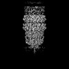
| Title | Top cap of bacteriophage Mycofy1 mature head (C5 symmetry) | |||||||||
 Components Components | Phage capsid-like C-terminal domain-containing protein | |||||||||
 Keywords Keywords | VIRAL PROTEIN / Mycobacterium / bacteriophage / prolate head / major capsid protein / VIRUS | |||||||||
| Function / homology | : / Phage capsid / Phage capsid family / virion component / Phage capsid-like C-terminal domain-containing protein Function and homology information Function and homology information | |||||||||
| Biological species |  Mycolicibacterium phage Mycofy1 (virus) Mycolicibacterium phage Mycofy1 (virus) | |||||||||
| Method | ELECTRON MICROSCOPY / single particle reconstruction / cryo EM / Resolution: 3.42 Å | |||||||||
 Authors Authors | Li, X. / Shao, Q. / Li, L. / Xie, L. / Ruan, Z. / Fang, Q. | |||||||||
| Funding support |  China, 2items China, 2items
| |||||||||
 Citation Citation | Journal: J Mol Biol / Year: 2025 Title: Cryo-EM Reveals Structural Diversity in Prolate-headed Mycobacteriophage Mycofy1. Authors: Xiangyun Li / Qianqian Shao / Lin Li / Linlin Xie / Zhiyang Ruan / Qianglin Fang / Abstract: Mycobacteriophages show promise in treating antibiotic-resistant mycobacterial infections. Here, we isolated Mycofy1, a mycobacteriophage, using M. smegmatis as a host. Cryo-EM analysis revealed that ...Mycobacteriophages show promise in treating antibiotic-resistant mycobacterial infections. Here, we isolated Mycofy1, a mycobacteriophage, using M. smegmatis as a host. Cryo-EM analysis revealed that Mycofy1 possesses a prolate head and a long non-contractile tail. We determined structures of its head, head-to-tail interface, terminator, and tail tube to resolutions of ∼3.5 Å. Unexpectedly, we identified two distinct types of prolate head structures, exhibiting a 36° relative rotation in the top cap region. Additionally, the head-to-tail interface demonstrated flexibility. Our structures provide high-resolution cryo-EM data of a mycobacteriophage with a prolate head, as well as detailed structural information of the head-to-tail interface and head-proximal tail region in this phage group. These findings advance our understanding of assembly mechanisms in tailed bacteriophages. | |||||||||
| History |
|
- Structure visualization
| Structure viewer | Molecule: MolmilJmol/JSmol |
|---|
- Downloads & links
-Download
| PDBx/mmCIF format | 9lw6.cif.gz | 2.6 MB | Display | PDBx/mmCIF format |
|---|---|---|---|---|
| PDB format | pdb9lw6.ent.gz | Display | PDB format | |
| PDBx/mmJSON format | 9lw6.json.gz | Tree view | PDBx/mmJSON format | |
| Others |  Other downloads Other downloads |
-Validation report
| Arichive directory | https://data.pdbj.org/pub/pdb/validation_reports/lw/9lw6ftp://data.pdbj.org/pub/pdb/validation_reports/lw/9lw6 | HTTPS FTP |
|---|
-Related structure data
| Related structure data | 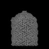 63432MC 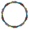 9lw7C 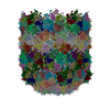 9lw8C 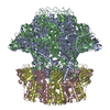 9lw9C 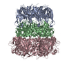 9lwaC M: map data used to model this data C: citing same article ( |
|---|---|
| Similar structure data |
-Links
 PDBj
PDBj

- Assembly
| Deposited unit | 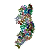
|
|---|---|
| 1 | x 5
|
| 2 |
|
| 3 | 
|
| Symmetry | Point symmetry: (Schoenflies symbol: C5 (5 fold cyclic)) |
-Components
| #1: Protein | Mass: 59777.262 Da / Num. of mol.: 54 / Source method: isolated from a natural source Details: Sequence reference for Mycolicibacterium phage Mycofy1 is not available at the time of biocuration. Current sequence reference is from UniProt id Q854Z2. Source: (natural) Mycolicibacterium phage Mycofy1 (virus) / References: UniProt: Q854Z2Has protein modification | N | |
|---|
-Experimental details
-Experiment
| Experiment | Method: ELECTRON MICROSCOPY |
|---|---|
| EM experiment | Aggregation state: PARTICLE / 3D reconstruction method: single particle reconstruction |
- Sample preparation
| Component | Name: Mycolicibacterium phage Mycofy1 / Type: VIRUS / Entity ID: all / Source: NATURAL |
|---|---|
| Molecular weight | Experimental value: NO |
| Source (natural) | Organism: Mycolicibacterium phage Mycofy1 (virus) |
| Details of virus | Empty: NO / Enveloped: NO / Isolate: STRAIN / Type: VIRION |
| Natural host | Organism: Mycolicibacterium smegmatis MC2 155 |
| Buffer solution | pH: 7.5 |
| Specimen | Embedding applied: NO / Shadowing applied: NO / Staining applied: NO / Vitrification applied: YES |
| Specimen support | Grid material: COPPER / Grid type: Quantifoil R2/1 |
| Vitrification | Instrument: FEI VITROBOT MARK IV / Cryogen name: ETHANE / Humidity: 100 % |
- Electron microscopy imaging
| Experimental equipment |  Model: Titan Krios / Image courtesy: FEI Company |
|---|---|
| Microscopy | Model: FEI TITAN KRIOS |
| Electron gun | Electron source:  FIELD EMISSION GUN / Accelerating voltage: 300 kV / Illumination mode: FLOOD BEAM FIELD EMISSION GUN / Accelerating voltage: 300 kV / Illumination mode: FLOOD BEAM |
| Electron lens | Mode: BRIGHT FIELD / Nominal magnification: 59000 X / Nominal defocus max: 3000 nm / Nominal defocus min: 1000 nm / C2 aperture diameter: 70 µm |
| Specimen holder | Specimen holder model: FEI TITAN KRIOS AUTOGRID HOLDER |
| Image recording | Average exposure time: 5.09 sec. / Electron dose: 25.7 e/Å2 / Film or detector model: FEI FALCON IV (4k x 4k) |
- Processing
| EM software |
| ||||||||||||||||||||||||||||
|---|---|---|---|---|---|---|---|---|---|---|---|---|---|---|---|---|---|---|---|---|---|---|---|---|---|---|---|---|---|
| CTF correction | Type: PHASE FLIPPING AND AMPLITUDE CORRECTION | ||||||||||||||||||||||||||||
| Particle selection | Num. of particles selected: 22074 / Details: Manual picking | ||||||||||||||||||||||||||||
| Symmetry | Point symmetry: C5 (5 fold cyclic) | ||||||||||||||||||||||||||||
| 3D reconstruction | Resolution: 3.42 Å / Resolution method: FSC 0.143 CUT-OFF / Num. of particles: 19765 / Symmetry type: POINT | ||||||||||||||||||||||||||||
| Refine LS restraints |
|
