 Movie
Movie Controller


+ Open data

- Basic information
| Entry | 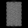 | |||||||||
|---|---|---|---|---|---|---|---|---|---|---|
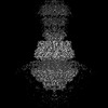
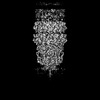
| Title | Midsection of bacteriophage Mycofy1 mature head (C5 symmetry) | |||||||||
 Map data Map data | ||||||||||
 Sample Sample |
| |||||||||
 Keywords Keywords | Mycobacterium / bacteriophage / prolate head / major capsid protein / VIRUS / VIRAL PROTEIN | |||||||||
| Function / homology | : / Phage capsid / Phage capsid family / virion component / Phage capsid-like C-terminal domain-containing protein Function and homology information Function and homology information | |||||||||
| Biological species |  Mycolicibacterium phage Mycofy1 (virus) Mycolicibacterium phage Mycofy1 (virus) | |||||||||
| Method | single particle reconstruction / cryo EM / Resolution: 3.52 Å | |||||||||
 Authors Authors | Li X / Shao Q / Li L / Xie L / Ruan Z / Fang Q | |||||||||
| Funding support |  China, 2 items China, 2 items
| |||||||||
 Citation Citation | Journal: J Mol Biol / Year: 2025 Title: Cryo-EM Reveals Structural Diversity in Prolate-headed Mycobacteriophage Mycofy1. Authors: Xiangyun Li / Qianqian Shao / Lin Li / Linlin Xie / Zhiyang Ruan / Qianglin Fang / Abstract: Mycobacteriophages show promise in treating antibiotic-resistant mycobacterial infections. Here, we isolated Mycofy1, a mycobacteriophage, using M. smegmatis as a host. Cryo-EM analysis revealed that ...Mycobacteriophages show promise in treating antibiotic-resistant mycobacterial infections. Here, we isolated Mycofy1, a mycobacteriophage, using M. smegmatis as a host. Cryo-EM analysis revealed that Mycofy1 possesses a prolate head and a long non-contractile tail. We determined structures of its head, head-to-tail interface, terminator, and tail tube to resolutions of ∼3.5 Å. Unexpectedly, we identified two distinct types of prolate head structures, exhibiting a 36° relative rotation in the top cap region. Additionally, the head-to-tail interface demonstrated flexibility. Our structures provide high-resolution cryo-EM data of a mycobacteriophage with a prolate head, as well as detailed structural information of the head-to-tail interface and head-proximal tail region in this phage group. These findings advance our understanding of assembly mechanisms in tailed bacteriophages. | |||||||||
| History |
|
- Structure visualization
| Supplemental images |
|---|

- Downloads & links
-EMDB archive
| Map data | emd_63433.map.gz | 440.7 MB | EMDB map data format | |
|---|---|---|---|---|
| Header (meta data) | emd-63433-v30.xmlemd-63433.xml | 17 KB 17 KB | Display Display | EMDB header |
| FSC (resolution estimation) | emd_63433_fsc.xml | 17.5 KB | Display | FSC data file |
| Images |  emd_63433.png emd_63433.png | 204.9 KB | ||
| Masks | emd_63433_msk_1.map | 476.8 MB | Mask map | |
| Filedesc metadata | emd-63433.cif.gz | 6 KB | ||
| Others | emd_63433_half_map_1.map.gzemd_63433_half_map_2.map.gz | 381.4 MB 381.3 MB | ||
| Archive directory |  http://ftp.pdbj.org/pub/emdb/structures/EMD-63433ftp://ftp.pdbj.org/pub/emdb/structures/EMD-63433 http://ftp.pdbj.org/pub/emdb/structures/EMD-63433ftp://ftp.pdbj.org/pub/emdb/structures/EMD-63433 | HTTPS FTP |
-Related structure data
| Related structure data |  9lw7MC 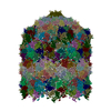 9lw6C 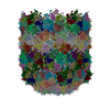 9lw8C 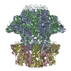 9lw9C 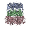 9lwaC M: atomic model generated by this map C: citing same article ( |
|---|---|
| Similar structure data |
-Links
| EMDB pages | EMDB (EBI/PDBe) / EMDataResource |
|---|---|
| Related items in Molecule of the Month |


-Map
| File | Download / File: emd_63433.map.gz / Format: CCP4 / Size: 476.8 MB / Type: IMAGE STORED AS FLOATING POINT NUMBER (4 BYTES) | ||||||||||||||||||||||||||||||||||||
|---|---|---|---|---|---|---|---|---|---|---|---|---|---|---|---|---|---|---|---|---|---|---|---|---|---|---|---|---|---|---|---|---|---|---|---|---|---|
| Projections & slices | Image control
Images are generated by Spider. | ||||||||||||||||||||||||||||||||||||
| Voxel size | X=Y=Z: 1.6464 Å | ||||||||||||||||||||||||||||||||||||
| Density |
| ||||||||||||||||||||||||||||||||||||
| Symmetry | Space group: 1 | ||||||||||||||||||||||||||||||||||||
| Details | EMDB XML:
|
 Z (Sec.)
 Y (Row.)
 X (Col.)


-Supplemental data
-Mask #1
| File | emd_63433_msk_1.map | ||||||||||||
|---|---|---|---|---|---|---|---|---|---|---|---|---|---|
| Projections & Slices |
| ||||||||||||
| Density Histograms |


-Half map: #2
| File | emd_63433_half_map_1.map | ||||||||||||
|---|---|---|---|---|---|---|---|---|---|---|---|---|---|
| Projections & Slices |
| ||||||||||||
| Density Histograms |


-Half map: #1
| File | emd_63433_half_map_2.map | ||||||||||||
|---|---|---|---|---|---|---|---|---|---|---|---|---|---|
| Projections & Slices |
| ||||||||||||
| Density Histograms |


- Sample components
-Entire : Mycolicibacterium phage Mycofy1
| Entire | Name: Mycolicibacterium phage Mycofy1 (virus) |
|---|---|
| Components |
|
-Supramolecule #1: Mycolicibacterium phage Mycofy1
| Supramolecule | Name: Mycolicibacterium phage Mycofy1 / type: virus / ID: 1 / Parent: 0 / Macromolecule list: all / NCBI-ID: 3349809 / Sci species name: Mycolicibacterium phage Mycofy1 / Virus type: VIRION / Virus isolate: STRAIN / Virus enveloped: No / Virus empty: No |
|---|---|
| Host (natural) | Organism:  Mycolicibacterium smegmatis MC2 155 (bacteria) Mycolicibacterium smegmatis MC2 155 (bacteria) |
-Macromolecule #1: Phage capsid-like C-terminal domain-containing protein
| Macromolecule | Name: Phage capsid-like C-terminal domain-containing protein type: protein_or_peptide / ID: 1 Details: Sequence reference for Mycolicibacterium phage Mycofy1 is not available at the time of biocuration. Current sequence reference is from UniProt id Q854Z2. Number of copies: 12 / Enantiomer: LEVO |
|---|---|
| Source (natural) | Organism: Mycolicibacterium phage Mycofy1 (virus) |
| Molecular weight | Theoretical: 59.777262 KDa |
| Sequence | String: MNTLDTLPVH PRTGLRAIGM GKRGPIWPVM GASDDHKDDA PTLTYSQARN RADEVHARME QIAELDKPTD EENEEFRALG AEFDSLVNH MSRLERAAEL ARVRSTHEQI GKPQSGGQRR MRVEAGSSQG GRGDYDRDAI LEPDSIEDCR FRDPWNLSEM R TFGRDAEE ...String: MNTLDTLPVH PRTGLRAIGM GKRGPIWPVM GASDDHKDDA PTLTYSQARN RADEVHARME QIAELDKPTD EENEEFRALG AEFDSLVNH MSRLERAAEL ARVRSTHEQI GKPQSGGQRR MRVEAGSSQG GRGDYDRDAI LEPDSIEDCR FRDPWNLSEM R TFGRDAEE VKGELRARAL SAIEKMQGAS DNVRAAATHI IERFDDEDST LARQCLATSS PAYLRAWSKM ARNPHAAILT EE EKRAINE VRAMGLTKAD GGYLVPFQLD PTVIITSNGS LNDIRRFARQ VVATGDVWHG VSSAAVQWSW DAEFEEVSDD SPE FGQPEI PVKKAQGFVP ISIEALQDEA NVTETVALLF AEGKDELEAV TLTTGTGQGN QPTGIVTALA GTAAEIAPVT AETF ALADV YAVYEQLAAR HRRQGAWLAN NLIYNKIRQF DTQGGAGLWT TIGNGEPSQL LGRPVGEAEA MDANWNTSAS ADNFV LLYG NFQNYVIADR IGMTVEFIPH LFGTNRRPNG SRGWFAYYRM GADVVNPNAF RLLNVETAS UniProtKB: Phage capsid-like C-terminal domain-containing protein |
-Experimental details
-Structure determination
| Method | cryo EM |
|---|---|
 Processing Processing | single particle reconstruction |
| Aggregation state | particle |
-Sample preparation
| Buffer | pH: 7.5 |
|---|---|
| Grid | Model: Quantifoil R2/1 / Material: COPPER / Support film - Material: CARBON / Support film - topology: CONTINUOUS / Pretreatment - Type: GLOW DISCHARGE |
| Vitrification | Cryogen name: ETHANE / Chamber humidity: 100 % / Instrument: FEI VITROBOT MARK IV |
- Electron microscopy
| Microscope | TFS KRIOS |
|---|---|
| Software | Name: EPU |
| Image recording | Film or detector model: FEI FALCON IV (4k x 4k) / Average exposure time: 5.09 sec. / Average electron dose: 25.7 e/Å2 |
| Electron beam | Acceleration voltage: 300 kV / Electron source:  FIELD EMISSION GUN FIELD EMISSION GUN |
| Electron optics | C2 aperture diameter: 70.0 µm / Illumination mode: FLOOD BEAM / Imaging mode: BRIGHT FIELD / Nominal defocus max: 3.0 µm / Nominal defocus min: 1.0 µm / Nominal magnification: 59000 |
| Sample stage | Specimen holder model: FEI TITAN KRIOS AUTOGRID HOLDER |
| Experimental equipment |  Model: Titan Krios / Image courtesy: FEI Company |

