 Movie
Movie Controller

[English] 日本語
 Yorodumi
- EMDB-63436: Bacteriophage Mycofy1 distal head-to-tail interface (C6 symmetry) -
+ Open data

- Basic information
| Entry | 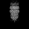 | |||||||||
|---|---|---|---|---|---|---|---|---|---|---|
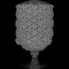
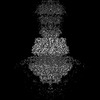
| Title | Bacteriophage Mycofy1 distal head-to-tail interface (C6 symmetry) | |||||||||
 Map data Map data | ||||||||||
 Sample Sample |
| |||||||||
 Keywords Keywords | Mycobacterium / bacteriophage / prolate head / head-to-tail interface / connector protein / terminator protein / tail tube protein / VIRUS / VIRAL PROTEIN | |||||||||
| Function / homology | Major tail protein / Head-to-tail stopper Function and homology information Function and homology information | |||||||||
| Biological species |  Mycolicibacterium phage Mycofy1 (virus) Mycolicibacterium phage Mycofy1 (virus) | |||||||||
| Method | single particle reconstruction / cryo EM / Resolution: 3.83 Å | |||||||||
 Authors Authors | Li X / Shao Q / Li L / Xie L / Ruan Z / Fang Q | |||||||||
| Funding support |  China, 2 items China, 2 items
| |||||||||
 Citation Citation | Journal: J Mol Biol / Year: 2025 Title: Cryo-EM Reveals Structural Diversity in Prolate-headed Mycobacteriophage Mycofy1. Authors: Xiangyun Li / Qianqian Shao / Lin Li / Linlin Xie / Zhiyang Ruan / Qianglin Fang / Abstract: Mycobacteriophages show promise in treating antibiotic-resistant mycobacterial infections. Here, we isolated Mycofy1, a mycobacteriophage, using M. smegmatis as a host. Cryo-EM analysis revealed that ...Mycobacteriophages show promise in treating antibiotic-resistant mycobacterial infections. Here, we isolated Mycofy1, a mycobacteriophage, using M. smegmatis as a host. Cryo-EM analysis revealed that Mycofy1 possesses a prolate head and a long non-contractile tail. We determined structures of its head, head-to-tail interface, terminator, and tail tube to resolutions of ∼3.5 Å. Unexpectedly, we identified two distinct types of prolate head structures, exhibiting a 36° relative rotation in the top cap region. Additionally, the head-to-tail interface demonstrated flexibility. Our structures provide high-resolution cryo-EM data of a mycobacteriophage with a prolate head, as well as detailed structural information of the head-to-tail interface and head-proximal tail region in this phage group. These findings advance our understanding of assembly mechanisms in tailed bacteriophages. | |||||||||
| History |
|
- Structure visualization
| Supplemental images |
|---|

- Downloads & links
-EMDB archive
| Map data | emd_63436.map.gz | 48.4 MB | EMDB map data format | |
|---|---|---|---|---|
| Header (meta data) | emd-63436-v30.xmlemd-63436.xml | 19.2 KB 19.2 KB | Display Display | EMDB header |
| FSC (resolution estimation) | emd_63436_fsc.xml | 8.5 KB | Display | FSC data file |
| Images |  emd_63436.png emd_63436.png | 91.5 KB | ||
| Masks | emd_63436_msk_1.map | 52.7 MB | Mask map | |
| Filedesc metadata | emd-63436.cif.gz | 6.1 KB | ||
| Others | emd_63436_half_map_1.map.gzemd_63436_half_map_2.map.gz | 39.6 MB 39.6 MB | ||
| Archive directory |  http://ftp.pdbj.org/pub/emdb/structures/EMD-63436ftp://ftp.pdbj.org/pub/emdb/structures/EMD-63436 http://ftp.pdbj.org/pub/emdb/structures/EMD-63436ftp://ftp.pdbj.org/pub/emdb/structures/EMD-63436 | HTTPS FTP |
-Related structure data
| Related structure data | 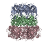 9lwaMC 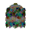 9lw6C 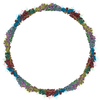 9lw7C 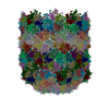 9lw8C 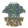 9lw9C M: atomic model generated by this map C: citing same article ( |
|---|---|
| Similar structure data |
-Links
| EMDB pages | EMDB (EBI/PDBe) / EMDataResource |
|---|
-Map
| File | Download / File: emd_63436.map.gz / Format: CCP4 / Size: 52.7 MB / Type: IMAGE STORED AS FLOATING POINT NUMBER (4 BYTES) | ||||||||||||||||||||||||||||||||||||
|---|---|---|---|---|---|---|---|---|---|---|---|---|---|---|---|---|---|---|---|---|---|---|---|---|---|---|---|---|---|---|---|---|---|---|---|---|---|
| Projections & slices | Image control
Images are generated by Spider. | ||||||||||||||||||||||||||||||||||||
| Voxel size | X=Y=Z: 1.372 Å | ||||||||||||||||||||||||||||||||||||
| Density |
| ||||||||||||||||||||||||||||||||||||
| Symmetry | Space group: 1 | ||||||||||||||||||||||||||||||||||||
| Details | EMDB XML:
|
 Z (Sec.)
 Y (Row.)
 X (Col.)


-Supplemental data
-Mask #1
| File | emd_63436_msk_1.map | ||||||||||||
|---|---|---|---|---|---|---|---|---|---|---|---|---|---|
| Projections & Slices |
| ||||||||||||
| Density Histograms |


-Half map: #1
| File | emd_63436_half_map_1.map | ||||||||||||
|---|---|---|---|---|---|---|---|---|---|---|---|---|---|
| Projections & Slices |
| ||||||||||||
| Density Histograms |


-Half map: #2
| File | emd_63436_half_map_2.map | ||||||||||||
|---|---|---|---|---|---|---|---|---|---|---|---|---|---|
| Projections & Slices |
| ||||||||||||
| Density Histograms |


- Sample components
-Entire : Mycolicibacterium phage Mycofy1
| Entire | Name: Mycolicibacterium phage Mycofy1 (virus) |
|---|---|
| Components |
|
-Supramolecule #1: Mycolicibacterium phage Mycofy1
| Supramolecule | Name: Mycolicibacterium phage Mycofy1 / type: virus / ID: 1 / Parent: 0 / Macromolecule list: all / NCBI-ID: 3349809 / Sci species name: Mycolicibacterium phage Mycofy1 / Virus type: VIRION / Virus isolate: STRAIN / Virus enveloped: No / Virus empty: No |
|---|---|
| Host (natural) | Organism:  Mycolicibacterium smegmatis MC2 155 (bacteria) Mycolicibacterium smegmatis MC2 155 (bacteria) |
-Macromolecule #1: Head-to-tail stopper
| Macromolecule | Name: Head-to-tail stopper / type: protein_or_peptide / ID: 1 Details: Sequence reference for Mycolicibacterium phage Mycofy1 is not available at the time of biocuration. Current sequence reference is from UniProt id Q854Y9. Number of copies: 1 / Enantiomer: LEVO |
|---|---|
| Source (natural) | Organism: Mycolicibacterium phage Mycofy1 (virus) |
| Molecular weight | Theoretical: 13.696388 KDa |
| Sequence | String: MSEEFGGQVV ALVSVAETGV PGWGGLKAKA RTTTRQPGVH FRPASSTETP DGQTNVATEV WKLTAPPTAA VLAAKPGGEL VYDGTEHPE SLDLDSAAGR AATFRVDGPI MPKYDLAGQV HHVTIMCKRQ AG UniProtKB: Head-to-tail stopper |
-Macromolecule #2: Terminator protein gp11
| Macromolecule | Name: Terminator protein gp11 / type: protein_or_peptide / ID: 2 / Number of copies: 1 / Enantiomer: LEVO |
|---|---|
| Source (natural) | Organism: Mycolicibacterium phage Mycofy1 (virus) |
| Molecular weight | Theoretical: 15.704601 KDa |
| Sequence | String: MTMADLHDQD APDEEDFVVC WMQPVMRTAV ERDIDAELPF CEVTRIDGAD DPEAGTDDPV IQLDFYALGA EAAKAAAKQG HRRMLFLFR NFPTVTLSDG TLADLDFGET LIKPSRMAFE HDQIVRYTAR YQLGTSYVAV P |
-Macromolecule #3: Major tail protein
| Macromolecule | Name: Major tail protein / type: protein_or_peptide / ID: 3 Details: Sequence reference for Mycolicibacterium phage Mycofy1 is not available at the time of biocuration. Current sequence reference is from UniProt id A0A0A7RVP8. Number of copies: 1 / Enantiomer: LEVO |
|---|---|
| Source (natural) | Organism: Mycolicibacterium phage Mycofy1 (virus) |
| Molecular weight | Theoretical: 29.029006 KDa |
| Sequence | String: MTQPLTGTTP AAGGYTTVDN RLQGGHRTGL IAVAFRDALG TSTNISPHNA NGSVRWSPLA QDGQLRDDLF AHRLENGVWV ENTNPNEGW YLAGAFGEGN GPSSRPSIDT DDQMIEQSNW PFESDITKQD EPFTFQALQN LYPAIQRLAN NLPLSDANGN P LVELPGEA ...String: MTQPLTGTTP AAGGYTTVDN RLQGGHRTGL IAVAFRDALG TSTNISPHNA NGSVRWSPLA QDGQLRDDLF AHRLENGVWV ENTNPNEGW YLAGAFGEGN GPSSRPSIDT DDQMIEQSNW PFESDITKQD EPFTFQALQN LYPAIQRLAN NLPLSDANGN P LVELPGEA DGFSQPVDAE KIGRQFLLYG IRKKEGRYLY EVDAYDLAYL NNKGERKFGK RGTAAELTFK PEPSGYFMAM VD GEYKPII KHTFIGGPAW DALAGDGS UniProtKB: Major tail protein |
-Experimental details
-Structure determination
| Method | cryo EM |
|---|---|
 Processing Processing | single particle reconstruction |
| Aggregation state | particle |
-Sample preparation
| Buffer | pH: 7.5 |
|---|---|
| Grid | Model: Quantifoil R2/1 / Material: COPPER / Support film - Material: CARBON / Support film - topology: CONTINUOUS / Pretreatment - Type: GLOW DISCHARGE |
| Vitrification | Cryogen name: ETHANE / Chamber humidity: 100 % / Instrument: FEI VITROBOT MARK IV |
- Electron microscopy
| Microscope | TFS KRIOS |
|---|---|
| Software | Name: EPU |
| Image recording | Film or detector model: FEI FALCON IV (4k x 4k) / Average exposure time: 5.09 sec. / Average electron dose: 25.7 e/Å2 |
| Electron beam | Acceleration voltage: 300 kV / Electron source:  FIELD EMISSION GUN FIELD EMISSION GUN |
| Electron optics | C2 aperture diameter: 70.0 µm / Illumination mode: FLOOD BEAM / Imaging mode: BRIGHT FIELD / Nominal defocus max: 3.0 µm / Nominal defocus min: 1.0 µm / Nominal magnification: 59000 |
| Sample stage | Specimen holder model: FEI TITAN KRIOS AUTOGRID HOLDER |
| Experimental equipment |  Model: Titan Krios / Image courtesy: FEI Company |

