Ministry of Education, Culture, Sports, Science and Technology (Japan)
日本
引用
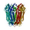
ジャーナル: Biochem Biophys Res Commun / 年: 2025 タイトル: Cryo-EM structure of the human Pannexin-3 channel. 著者: Taiichi Tsuyama / Ryuga Teramura / Kaoru Mitsuoka / Jun-Ichi Kishikawa / Ken Yokoyama / 要旨: Pannexin-3 (PANX3) is a member of the pannexin family of large-pore, ATP-permeable channels conserved across vertebrates. PANX3 contributes to various developmental and pathophysiological processes ...Pannexin-3 (PANX3) is a member of the pannexin family of large-pore, ATP-permeable channels conserved across vertebrates. PANX3 contributes to various developmental and pathophysiological processes by permeating ATP and Ca ions; however, the structural basis of PANX3 channel function remains unclear. Here, we present the cryo-EM structure of human PANX3 at 2.9-3.2 Å. The PANX3 channel is heptameric and forms a transmembrane pore along the central symmetric axis. The narrowest constriction of the pore is composed of an isoleucine ring located in the extracellular region, and its size is comparable to that of other pannexins. A structural variability analysis revealed prominent structural dynamics in intracellular regions. Our structural studies provide a foundation for understanding the detailed properties of pannexin channels.
 ムービー
ムービー コントローラー


 データを開く
 基本情報
基本情報 要素
要素 キーワード
キーワード 機能・相同性情報
 Homo sapiens (ヒト)
 データ登録者
 日本, 4件
  引用
引用 構造の表示
構造の表示 ダウンロードとリンク
 その他のダウンロード
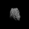
 PDBj
PDBj 集合体
集合体
 試料調製
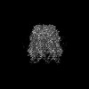
試料調製 電子顕微鏡撮影
 FIELD EMISSION GUN / 加速電圧: 300 kV / 照射モード: FLOOD BEAM
 解析
解析