 Movie
Movie Controller


+ Open data

- Basic information
| Entry | 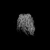 | |||||||||||||||
|---|---|---|---|---|---|---|---|---|---|---|---|---|---|---|---|---|
| Title | Cryo-EM structure of human pannexin-3 protomer | |||||||||||||||
 Map data Map data | ||||||||||||||||
 Sample Sample |
| |||||||||||||||
 Keywords Keywords | Pannexin / Innexin / TRANSPORT PROTEIN | |||||||||||||||
| Function / homology |  Function and homology information Function and homology informationgap junction hemi-channel activity / positive regulation of interleukin-1 production / wide pore channel activity / gap junction / monoatomic cation transport / osteoblast differentiation / calcium channel activity / cell-cell signaling / endoplasmic reticulum membrane / structural molecule activity / plasma membrane Similarity search - Function | |||||||||||||||
| Biological species |  Homo sapiens (human) Homo sapiens (human) | |||||||||||||||
| Method | single particle reconstruction / cryo EM / Resolution: 3.2 Å | |||||||||||||||
 Authors Authors | Tsuyama T / Yokoyama K | |||||||||||||||
| Funding support |  Japan, 4 items Japan, 4 items
| |||||||||||||||
 Citation Citation | Journal: Biochem Biophys Res Commun / Year: 2025 Title: Cryo-EM structure of the human Pannexin-3 channel. Authors: Taiichi Tsuyama / Ryuga Teramura / Kaoru Mitsuoka / Jun-Ichi Kishikawa / Ken Yokoyama / Abstract: Pannexin-3 (PANX3) is a member of the pannexin family of large-pore, ATP-permeable channels conserved across vertebrates. PANX3 contributes to various developmental and pathophysiological processes ...Pannexin-3 (PANX3) is a member of the pannexin family of large-pore, ATP-permeable channels conserved across vertebrates. PANX3 contributes to various developmental and pathophysiological processes by permeating ATP and Ca ions; however, the structural basis of PANX3 channel function remains unclear. Here, we present the cryo-EM structure of human PANX3 at 2.9-3.2 Å. The PANX3 channel is heptameric and forms a transmembrane pore along the central symmetric axis. The narrowest constriction of the pore is composed of an isoleucine ring located in the extracellular region, and its size is comparable to that of other pannexins. A structural variability analysis revealed prominent structural dynamics in intracellular regions. Our structural studies provide a foundation for understanding the detailed properties of pannexin channels. | |||||||||||||||
| History |
|
- Structure visualization
| Supplemental images |
|---|

- Downloads & links
-EMDB archive
| Map data | emd_62478.map.gz | 97 MB | EMDB map data format | |
|---|---|---|---|---|
| Header (meta data) | emd-62478-v30.xmlemd-62478.xml | 16.7 KB 16.7 KB | Display Display | EMDB header |
| FSC (resolution estimation) | emd_62478_fsc.xml | 9.9 KB | Display | FSC data file |
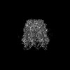
| Images |  emd_62478.png emd_62478.png | 133.8 KB | ||
| Masks | emd_62478_msk_1.map | 103 MB | Mask map | |
| Filedesc metadata | emd-62478.cif.gz | 5.9 KB | ||
| Others | emd_62478_half_map_1.map.gzemd_62478_half_map_2.map.gz | 95.4 MB 95.4 MB | ||
| Archive directory |  http://ftp.pdbj.org/pub/emdb/structures/EMD-62478ftp://ftp.pdbj.org/pub/emdb/structures/EMD-62478 http://ftp.pdbj.org/pub/emdb/structures/EMD-62478ftp://ftp.pdbj.org/pub/emdb/structures/EMD-62478 | HTTPS FTP |
-Related structure data
| Related structure data | 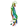 9komMC 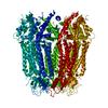 9krgC M: atomic model generated by this map C: citing same article ( |
|---|---|
| Similar structure data |
-Links
| EMDB pages | EMDB (EBI/PDBe) / EMDataResource |
|---|
-Map
| File | Download / File: emd_62478.map.gz / Format: CCP4 / Size: 103 MB / Type: IMAGE STORED AS FLOATING POINT NUMBER (4 BYTES) | ||||||||||||||||||||||||||||||||||||
|---|---|---|---|---|---|---|---|---|---|---|---|---|---|---|---|---|---|---|---|---|---|---|---|---|---|---|---|---|---|---|---|---|---|---|---|---|---|
| Projections & slices | Image control
Images are generated by Spider. | ||||||||||||||||||||||||||||||||||||
| Voxel size | X=Y=Z: 0.84 Å | ||||||||||||||||||||||||||||||||||||
| Density |
| ||||||||||||||||||||||||||||||||||||
| Symmetry | Space group: 1 | ||||||||||||||||||||||||||||||||||||
| Details | EMDB XML:
|
 Z (Sec.)
 Y (Row.)
 X (Col.)


-Supplemental data
-Mask #1
| File | emd_62478_msk_1.map | ||||||||||||
|---|---|---|---|---|---|---|---|---|---|---|---|---|---|
| Projections & Slices |
| ||||||||||||
| Density Histograms |


-Half map: #2
| File | emd_62478_half_map_1.map | ||||||||||||
|---|---|---|---|---|---|---|---|---|---|---|---|---|---|
| Projections & Slices |
| ||||||||||||
| Density Histograms |


-Half map: #1
| File | emd_62478_half_map_2.map | ||||||||||||
|---|---|---|---|---|---|---|---|---|---|---|---|---|---|
| Projections & Slices |
| ||||||||||||
| Density Histograms |


- Sample components
-Entire : Pannexin-3 protomer
| Entire | Name: Pannexin-3 protomer |
|---|---|
| Components |
|
-Supramolecule #1: Pannexin-3 protomer
| Supramolecule | Name: Pannexin-3 protomer / type: complex / ID: 1 / Parent: 0 / Macromolecule list: all |
|---|---|
| Source (natural) | Organism: Homo sapiens (human) |
-Macromolecule #1: Pannexin-3
| Macromolecule | Name: Pannexin-3 / type: protein_or_peptide / ID: 1 / Number of copies: 1 / Enantiomer: LEVO |
|---|---|
| Source (natural) | Organism: Homo sapiens (human) |
| Molecular weight | Theoretical: 45.965449 KDa |
| Recombinant expression | Organism: Homo sapiens (human) |
| Sequence | String: MSLAHTAAEY MLSDALLPDR RGPRLKGLRL ELPLDRIVKF VAVGSPLLLM SLAFAQEFSS GSPISCFSPS NFSIRQAAYV DSSCWDSLL HHKQDGPGQD KMKSLWPHKA LPYSLLALAL LMYLPVLLWQ YAAVPALSSD LLFIISELDK SYNRSIRLVQ H MLKIRQKS ...String: MSLAHTAAEY MLSDALLPDR RGPRLKGLRL ELPLDRIVKF VAVGSPLLLM SLAFAQEFSS GSPISCFSPS NFSIRQAAYV DSSCWDSLL HHKQDGPGQD KMKSLWPHKA LPYSLLALAL LMYLPVLLWQ YAAVPALSSD LLFIISELDK SYNRSIRLVQ H MLKIRQKS SDPYVFWNEL EKARKERYFE FPLLERYLAC KQRSHSLVAT YLLRNSLLLI FTSATYLYLG HFHLDVFFQE EF SCSIKTG LLSDETHVPN LITCRLTSLS IFQIVSLSSV AIYTILVPVI IYNLTRLCRW DKRLLSVYEM LPAFDLLSRK MLG CPINDL NVILLFLRAN ISELISFSWL SVLCVLKDTT TQKHNIDTVV DFMTLLAGLE PSKPKHLTNS ACDEHPGSGG SGHH HHHH UniProtKB: Pannexin-3 |
-Experimental details
-Structure determination
| Method | cryo EM |
|---|---|
 Processing Processing | single particle reconstruction |
| Aggregation state | particle |
-Sample preparation
| Concentration | 2 mg/mL | |||||||||
|---|---|---|---|---|---|---|---|---|---|---|
| Buffer | pH: 8 Component:
| |||||||||
| Grid | Model: Quantifoil R1.2/1.3 / Material: COPPER / Mesh: 300 / Pretreatment - Type: GLOW DISCHARGE / Pretreatment - Time: 60 sec. | |||||||||
| Vitrification | Cryogen name: ETHANE / Chamber humidity: 100 % / Chamber temperature: 277 K / Instrument: FEI VITROBOT MARK IV |
- Electron microscopy
| Microscope | TFS KRIOS |
|---|---|
| Image recording | Film or detector model: GATAN K3 (6k x 4k) / Number grids imaged: 1 / Average electron dose: 50.0 e/Å2 |
| Electron beam | Acceleration voltage: 300 kV / Electron source:  FIELD EMISSION GUN FIELD EMISSION GUN |
| Electron optics | Illumination mode: FLOOD BEAM / Imaging mode: BRIGHT FIELD / Cs: 2.7 mm / Nominal defocus max: 2.2 µm / Nominal defocus min: 1.0 µm |
| Experimental equipment |  Model: Titan Krios / Image courtesy: FEI Company |
+Image processing

-Atomic model buiding 1
| Initial model | Chain - Source name: Other / Chain - Initial model type: in silico model Details: ModelAngelo model generated with the full-length amino acid sequences |
|---|---|
| Refinement | Protocol: AB INITIO MODEL |
| Output model | PDB-9kom: |
