 Movie
Movie Controller


+ Open data

- Basic information
| Entry | Database: PDB / ID: 9kjr | ||||||
|---|---|---|---|---|---|---|---|
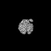
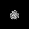
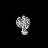
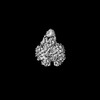
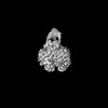
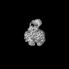
| Title | The cryo-EM structure of human PNPase in the open conformation | ||||||
 Components Components | Polyribonucleotide nucleotidyltransferase 1, mitochondrial | ||||||
 Keywords Keywords | RNA BINDING PROTEIN / 3'-to-5' exoribonuclease / RNA degradation / RNA import / mitochondria | ||||||
| Function / homology |  Function and homology information Function and homology informationRNA import into mitochondrion / mitochondrial mRNA polyadenylation / positive regulation of miRNA catabolic process / mitochondrial degradosome / mitochondrial mRNA catabolic process / positive regulation of mitochondrial RNA catabolic process / mitochondrial RNA 5'-end processing / mitochondrial RNA 3'-end processing / poly(G) binding / Mitochondrial RNA degradation ...RNA import into mitochondrion / mitochondrial mRNA polyadenylation / positive regulation of miRNA catabolic process / mitochondrial degradosome / mitochondrial mRNA catabolic process / positive regulation of mitochondrial RNA catabolic process / mitochondrial RNA 5'-end processing / mitochondrial RNA 3'-end processing / poly(G) binding / Mitochondrial RNA degradation / polyribonucleotide nucleotidyltransferase / polyribonucleotide nucleotidyltransferase activity / nuclear polyadenylation-dependent mRNA catabolic process / mitochondrial RNA catabolic process / positive regulation of mRNA catabolic process / regulation of cellular senescence / rRNA import into mitochondrion / regulation of cellular respiration / RNA catabolic process / response to growth hormone / miRNA binding / poly(U) RNA binding / protein homotrimerization / mRNA catabolic process / response to cAMP / cellular response to interferon-beta / liver regeneration / mitochondrion organization / protein homooligomerization / mitochondrial intermembrane space / mRNA processing / cellular response to oxidative stress / 3'-5'-RNA exonuclease activity / ribosome / mitochondrial matrix / endoplasmic reticulum membrane / mitochondrion / RNA binding / identical protein binding / cytoplasm / cytosol Similarity search - Function | ||||||
| Biological species |  Homo sapiens (human) Homo sapiens (human) | ||||||
| Method | ELECTRON MICROSCOPY / single particle reconstruction / cryo EM / Resolution: 3.86 Å | ||||||
 Authors Authors | Li, Y.C. / Yuan, H.S. | ||||||
| Funding support |  Taiwan, 1items Taiwan, 1items
| ||||||
 Citation Citation | Journal: Nucleic Acids Res / Year: 2025 Title: Structural insights into human PNPase in health and disease. Authors: Yi-Ching Li / Chun-Hsiung Wang / Malay Patra / Yi-Ping Chen / Wei-Zen Yang / Hanna S Yuan / Abstract: Human polynucleotide phosphorylase (hPNPase) is a 3'-to-5' exoribonuclease located in mitochondria, where it plays crucial roles in RNA degradation and RNA import. Mutations in hPNPase can impair ...Human polynucleotide phosphorylase (hPNPase) is a 3'-to-5' exoribonuclease located in mitochondria, where it plays crucial roles in RNA degradation and RNA import. Mutations in hPNPase can impair these functions, leading to various mitochondrial dysfunctions and diseases. However, the mechanisms by which hPNPase switches between its roles as an RNA-degrading enzyme and an RNA carrier, as well as how disease-associated mutations may affect these distinct functions, remain unclear. In this study, we present cryo-electron microscopy structures of hPNPase, highlighting the flexibility of its S1 domains, which cap the ring-like RNA-degradation chamber and shift between two distinctive open and closed conformations. We further demonstrate by small-angle X-ray scattering and biochemical analyses that the disease-associated mutations P467S and G499R impair hPNPase's stem-loop RNA-binding and degradation activities by limiting the S1 domain's ability to transition from an open to closed state. Conversely, the D713Y mutation, located within the S1 domain, does not affect the RNA-binding affinity of hPNPase, but diminishes its interaction with Suv3 helicase for cooperative degradation of structured RNA. Collectively, these findings underscore the critical role of S1 domain mobility in capturing structured RNA for degradation and import, as well as its involvement in mitochondrial degradosome assembly. Our study thereby reveals the molecular mechanism of hPNPase in RNA binding and degradation, and the multiple molecular defects that could be induced by disease-linked mutations in hPNPase. | ||||||
| History |
|
- Structure visualization
| Structure viewer | Molecule: MolmilJmol/JSmol |
|---|
- Downloads & links
-Download
| PDBx/mmCIF format | 9kjr.cif.gz | 358.3 KB | Display | PDBx/mmCIF format |
|---|---|---|---|---|
| PDB format | pdb9kjr.ent.gz | 294.1 KB | Display | PDB format |
| PDBx/mmJSON format | 9kjr.json.gz | Tree view | PDBx/mmJSON format | |
| Others |  Other downloads Other downloads |
-Validation report
| Arichive directory | https://data.pdbj.org/pub/pdb/validation_reports/kj/9kjrftp://data.pdbj.org/pub/pdb/validation_reports/kj/9kjr | HTTPS FTP |
|---|
-Related structure data
| Related structure data |  62372MC 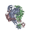 9kjtC C: citing same article ( M: map data used to model this data |
|---|---|
| Similar structure data |
-Links
 PDBj
PDBj


- Assembly
| Deposited unit | 
|
|---|---|
| 1 |
|
-Components
| #1: Protein | Mass: 78007.211 Da / Num. of mol.: 3 Source method: isolated from a genetically manipulated source Source: (gene. exp.) Homo sapiens (human) / Gene: PNPT1, PNPASE / Production host:  References: UniProt: Q8TCS8, polyribonucleotide nucleotidyltransferase Has protein modification | N | |
|---|
-Experimental details
-Experiment
| Experiment | Method: ELECTRON MICROSCOPY |
|---|---|
| EM experiment | Aggregation state: PARTICLE / 3D reconstruction method: single particle reconstruction |
- Sample preparation
| Component | Name: Human PNPase in open form / Type: COMPLEX / Entity ID: all / Source: RECOMBINANT |
|---|---|
| Source (natural) | Organism: Homo sapiens (human) |
| Source (recombinant) | Organism: |
| Buffer solution | pH: 7.4 |
| Specimen | Embedding applied: NO / Shadowing applied: NO / Staining applied: NO / Vitrification applied: YES |
| Vitrification | Cryogen name: ETHANE |
- Electron microscopy imaging
| Experimental equipment |  Model: Titan Krios / Image courtesy: FEI Company |
|---|---|
| Microscopy | Model: TFS KRIOS |
| Electron gun | Electron source:  FIELD EMISSION GUN / Accelerating voltage: 300 kV / Illumination mode: FLOOD BEAM FIELD EMISSION GUN / Accelerating voltage: 300 kV / Illumination mode: FLOOD BEAM |
| Electron lens | Mode: BRIGHT FIELD / Nominal defocus max: 2000 nm / Nominal defocus min: 1600 nm |
| Image recording | Electron dose: 50 e/Å2 / Film or detector model: GATAN K3 (6k x 4k) |
- Processing
| EM software | Name: PHENIX / Version: 1.20.1_4487 / Category: model refinement | ||||||||||||||||||||||||
|---|---|---|---|---|---|---|---|---|---|---|---|---|---|---|---|---|---|---|---|---|---|---|---|---|---|
| CTF correction | Type: PHASE FLIPPING AND AMPLITUDE CORRECTION | ||||||||||||||||||||||||
| 3D reconstruction | Resolution: 3.86 Å / Resolution method: FSC 0.143 CUT-OFF / Num. of particles: 91100 / Symmetry type: POINT | ||||||||||||||||||||||||
| Refinement | Highest resolution: 3.86 Å Stereochemistry target values: REAL-SPACE (WEIGHTED MAP SUM AT ATOM CENTERS) | ||||||||||||||||||||||||
| Refine LS restraints |
|
