 Movie
Movie Controller


+ Open data

- Basic information
| Entry | Database: PDB / ID: 9jqn | |||||||||||||||||||||
|---|---|---|---|---|---|---|---|---|---|---|---|---|---|---|---|---|---|---|---|---|---|---|
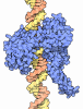
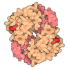
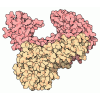
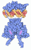
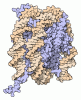
| Title | CryoEM structure of mouse RAG SEC-2DNA | |||||||||||||||||||||
 Components Components |
| |||||||||||||||||||||
 Keywords Keywords | DNA BINDING PROTEIN/DNA / V(D)J recombination / RAG / PHD / Transposition / DNA BINDING PROTEIN / DNA BINDING PROTEIN-DNA complex | |||||||||||||||||||||
| Function / homology |  Function and homology information Function and homology informationB cell homeostatic proliferation / T cell lineage commitment / mature B cell differentiation involved in immune response / B cell lineage commitment / negative regulation of T cell differentiation in thymus / DNA recombinase complex / endodeoxyribonuclease complex / negative regulation of T cell apoptotic process / positive regulation of organ growth / pre-B cell allelic exclusion ...B cell homeostatic proliferation / T cell lineage commitment / mature B cell differentiation involved in immune response / B cell lineage commitment / negative regulation of T cell differentiation in thymus / DNA recombinase complex / endodeoxyribonuclease complex / negative regulation of T cell apoptotic process / positive regulation of organ growth / pre-B cell allelic exclusion / V(D)J recombination / negative regulation of thymocyte apoptotic process / regulation of T cell differentiation / phosphatidylinositol-3,4-bisphosphate binding / T cell homeostasis / regulation of behavioral fear response / histone H3K4me3 reader activity / phosphatidylinositol-3,5-bisphosphate binding / positive regulation of T cell differentiation / thymus development / phosphatidylinositol-3,4,5-trisphosphate binding / T cell differentiation / T cell differentiation in thymus / protein autoubiquitination / B cell differentiation / phosphatidylinositol-4,5-bisphosphate binding / visual learning / phosphatidylinositol binding / RING-type E3 ubiquitin transferase / ubiquitin-protein transferase activity / ubiquitin protein ligase activity / endonuclease activity / DNA recombination / histone binding / chromatin organization / sequence-specific DNA binding / Hydrolases; Acting on ester bonds / adaptive immune response / defense response to bacterium / hydrolase activity / chromatin binding / protein homodimerization activity / nucleoplasm / zinc ion binding / metal ion binding / identical protein binding / nucleus Similarity search - Function | |||||||||||||||||||||
| Biological species |   Mus musculus (house mouse) Mus musculus (house mouse)  Escherichia coli (E. coli) Escherichia coli (E. coli) | |||||||||||||||||||||
| Method | ELECTRON MICROSCOPY / single particle reconstruction / cryo EM / Resolution: 3.03 Å | |||||||||||||||||||||
 Authors Authors | Chen, X. / Yao, L. / Yang, W. / Gellert, M. | |||||||||||||||||||||
| Funding support |  United States, United States,  China, 2items China, 2items
| |||||||||||||||||||||
 Citation Citation | Journal: Proc Natl Acad Sci U S A / Year: 2025 Title: How RAG1/2 evolved from ancestral transposases to initiate V(D)J recombination without transposition. Authors: Xuemin Chen / Liangrui Yao / Wenwen Li / Shanshan Ma / Xingyun Yuan / Yang Yang / Yuan Yuan / Yumei Liu / Lan Liu / Huaibin Wang / Martin Gellert / Wei Yang / Abstract: The recombination activating genes 1 and 2 (RAG1/2) recombinase, which initiates V(D)J recombination in jawed vertebrates, evolved from RNaseH-like transposases such as Transib and ProtoRAG. However, ...The recombination activating genes 1 and 2 (RAG1/2) recombinase, which initiates V(D)J recombination in jawed vertebrates, evolved from RNaseH-like transposases such as Transib and ProtoRAG. However, its postcleavage transposase activity is strictly suppressed. Previous structural studies have focused only on the conserved core domains of RAG1/2, leaving the regulatory mechanisms of the noncore regions unclear. To investigate how RAG1/2 suppresses transposition and regulates DNA cleavage, we determined cryo-electron microscopy (cryo-EM) structures of nearly full-length RAG1/2 complexed with cleaved recombination signal sequences (RSS) in a signal-end complex (SEC) at resolutions up to 2.95 Å. Two key structures, SEC-0 and SEC-Plant Homeodomain (PHD), reveal distinct regulatory roles of RAG2, which is absent in Transib transposase. SEC-0 displays a closed conformation, revealing that the core RAG2 facilitates sequential DNA cleavage by stabilizing the RSS-cleaved states in a "spring-loaded" mechanism. SEC-PHD reveals how RAG2's noncore PHD and Acidic Hinge (AH), which are absent in ProtoRAG, inhibit target DNA binding in transposition. Histone H3K4me3, which recruits RAG1/2 to RSS sites, does not influence RAG1/2 binding to V, D, or J gene segments bordered by RSS. In contrast, the suppressed transposition can be activated by H3K4me3 peptides that dislodge the inhibitory PHD. To achieve this derepression in vivo, however, would require an unlikely close placement of two nucleosomes flanking a target DNA bent by nearly 180°. Our structural and biochemical results elucidate how RAG1 has acquired RAG2 and utilizes its core and noncore domains to enhance V(D)J recombination and suppress transposition. | |||||||||||||||||||||
| History |
|
- Structure visualization
| Structure viewer | Molecule: MolmilJmol/JSmol |
|---|
- Downloads & links
-Download
| PDBx/mmCIF format | 9jqn.cif.gz | 399.9 KB | Display | PDBx/mmCIF format |
|---|---|---|---|---|
| PDB format | pdb9jqn.ent.gz | 300.9 KB | Display | PDB format |
| PDBx/mmJSON format | 9jqn.json.gz | Tree view | PDBx/mmJSON format | |
| Others |  Other downloads Other downloads |
-Validation report
| Arichive directory | https://data.pdbj.org/pub/pdb/validation_reports/jq/9jqnftp://data.pdbj.org/pub/pdb/validation_reports/jq/9jqn | HTTPS FTP |
|---|
-Related structure data
| Related structure data |  61730MC  9jpuC  9jpxC  9jtsC  9jtuC C: citing same article ( M: map data used to model this data |
|---|---|
| Similar structure data |


-Links
 PDBj
PDBj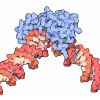
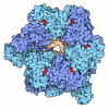

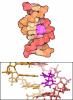


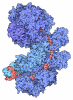

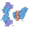
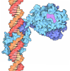
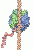
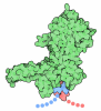


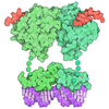






- Assembly
| Deposited unit | 
|
|---|---|
| 1 |
|
-Components
-V(D)J recombination-activating protein ... , 2 types, 4 molecules ACBD
| #1: Protein | Mass: 119389.352 Da / Num. of mol.: 2 Source method: isolated from a genetically manipulated source Source: (gene. exp.) Mus musculus (house mouse) / Gene: Rag1 / Production host:  Homo sapiens (human) Homo sapiens (human)References: UniProt: P15919, Hydrolases; Acting on ester bonds, RING-type E3 ubiquitin transferase #2: Protein | Mass: 59138.410 Da / Num. of mol.: 2 Source method: isolated from a genetically manipulated source Source: (gene. exp.) Mus musculus (house mouse) / Gene: Rag2, Rag-2 / Production host: Homo sapiens (human) / References: UniProt: P21784 |
|---|
-DNA chain , 6 types, 8 molecules GLMFHEIJ
| #3: DNA chain | Mass: 4574.982 Da / Num. of mol.: 1 / Source method: obtained synthetically / Source: (synth.) Escherichia coli (E. coli) | ||
|---|---|---|---|
| #4: DNA chain | Mass: 4562.988 Da / Num. of mol.: 1 / Source method: obtained synthetically / Source: (synth.) Escherichia coli (E. coli) | ||
| #5: DNA chain | Mass: 4602.024 Da / Num. of mol.: 1 / Source method: obtained synthetically / Source: (synth.) Escherichia coli (E. coli) | ||
| #6: DNA chain | Mass: 4615.995 Da / Num. of mol.: 1 / Source method: obtained synthetically / Source: (synth.) Escherichia coli (E. coli) | ||
| #7: DNA chain | Mass: 3945.589 Da / Num. of mol.: 2 / Source method: obtained synthetically / Source: (synth.) Escherichia coli (E. coli)#8: DNA chain | Mass: 3998.595 Da / Num. of mol.: 2 / Source method: obtained synthetically / Source: (synth.) Escherichia coli (E. coli) |
-Non-polymers , 2 types, 4 molecules 

| #9: Chemical |  Mass: 40.078 Da / Num. of mol.: 2 / Source method: obtained synthetically / Formula: Ca Mass: 40.078 Da / Num. of mol.: 2 / Source method: obtained synthetically / Formula: Ca#10: Chemical |  Mass: 65.409 Da / Num. of mol.: 2 / Source method: obtained synthetically / Formula: Zn Mass: 65.409 Da / Num. of mol.: 2 / Source method: obtained synthetically / Formula: Zn |
|---|
-Details
| Has ligand of interest | N |
|---|---|
| Has protein modification | N |
-Experimental details
-Experiment
| Experiment | Method: ELECTRON MICROSCOPY |
|---|---|
| EM experiment | Aggregation state: PARTICLE / 3D reconstruction method: single particle reconstruction |
- Sample preparation
| Component | Name: mouse RAG SEC / Type: COMPLEX / Entity ID: #1-#8 / Source: MULTIPLE SOURCES |
|---|---|
| Source (natural) | Organism: Mus musculus (house mouse) |
| Source (recombinant) | Organism: Homo sapiens (human) |
| Buffer solution | pH: 7.4 |
| Specimen | Embedding applied: NO / Shadowing applied: NO / Staining applied: NO / Vitrification applied: YES |
| Vitrification | Cryogen name: ETHANE |
- Electron microscopy imaging
| Experimental equipment |  Model: Titan Krios / Image courtesy: FEI Company |
|---|---|
| Microscopy | Model: TFS KRIOS |
| Electron gun | Electron source:  FIELD EMISSION GUN / Accelerating voltage: 300 kV / Illumination mode: FLOOD BEAM FIELD EMISSION GUN / Accelerating voltage: 300 kV / Illumination mode: FLOOD BEAM |
| Electron lens | Mode: BRIGHT FIELD / Nominal defocus max: 3000 nm / Nominal defocus min: 800 nm |
| Image recording | Electron dose: 70 e/Å2 / Film or detector model: GATAN K2 SUMMIT (4k x 4k) |
- Processing
| EM software | Name: PHENIX / Category: model refinement | ||||||||||||||||||||||||
|---|---|---|---|---|---|---|---|---|---|---|---|---|---|---|---|---|---|---|---|---|---|---|---|---|---|
| CTF correction | Details: cryoSPARC / Type: PHASE FLIPPING AND AMPLITUDE CORRECTION | ||||||||||||||||||||||||
| 3D reconstruction | Resolution: 3.03 Å / Resolution method: FSC 0.143 CUT-OFF / Num. of particles: 144322 / Symmetry type: POINT | ||||||||||||||||||||||||
| Refine LS restraints |
|
