 Movie
Movie Controller

[English] 日本語
 Yorodumi
- PDB-9jlu: GH57 family amylopullulanase from Aquifex aeolicus D352N mutant c... -
+ Open data

- Basic information
| Entry | Database: PDB / ID: 9jlu | ||||||
|---|---|---|---|---|---|---|---|
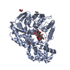
| Title | GH57 family amylopullulanase from Aquifex aeolicus D352N mutant complex with beta-cyclodextrin | ||||||
 Components Components | Glycoside hydrolase family 57 N-terminal domain-containing protein | ||||||
 Keywords Keywords | HYDROLASE / GH57 family / amylopullulanase / Aquifex aeolicus | ||||||
| Function / homology | : / Glycoside hydrolase family 57, N-terminal domain / Glycosyl hydrolase family 57 / Glycoside hydrolase 38, N-terminal domain superfamily / Glycoside hydrolase/deacetylase, beta/alpha-barrel / catalytic activity / carbohydrate metabolic process / beta-cyclodextrin / Glycoside hydrolase family 57 N-terminal domain-containing protein Function and homology information Function and homology information | ||||||
| Biological species |   Aquifex aeolicus VF5 (bacteria) Aquifex aeolicus VF5 (bacteria) | ||||||
| Method |  X-RAY DIFFRACTION / SYNCHROTRON / MOLECULAR REPLACEMENT / Resolution: 1.88 Å X-RAY DIFFRACTION / SYNCHROTRON / MOLECULAR REPLACEMENT / Resolution: 1.88 Å | ||||||
 Authors Authors | Zhu, Z.M. / Wang, W.W. / Li, M.J. / Xu, Q. / Zhou, H. / Huang, L.Q. / Wang, Q.S. / Yu, F. | ||||||
| Funding support | 1items
| ||||||
 Citation Citation | Journal: J.Struct.Biol. / Year: 2025 Title: Mechanistic insights into cyclodextrins as substrates and inhibitors of GH57 family amylopullulanase from Aquifex aeolicus. Authors: Zhu, Z. / Li, M. / Xu, Q. / Huang, L. / Zhou, H. / Wang, W. / Wang, Q. / Yu, F. | ||||||
| History |
|
- Structure visualization
| Structure viewer | Molecule: MolmilJmol/JSmol |
|---|
- Downloads & links
-Download
| PDBx/mmCIF format | 9jlu.cif.gz | 218.1 KB | Display | PDBx/mmCIF format |
|---|---|---|---|---|
| PDB format | pdb9jlu.ent.gz | 174.3 KB | Display | PDB format |
| PDBx/mmJSON format | 9jlu.json.gz | Tree view | PDBx/mmJSON format | |
| Others |  Other downloads Other downloads |
-Validation report
| Arichive directory | https://data.pdbj.org/pub/pdb/validation_reports/jl/9jluftp://data.pdbj.org/pub/pdb/validation_reports/jl/9jlu | HTTPS FTP |
|---|
-Related structure data


-Links
 PDBj
PDBj

- Assembly
| Deposited unit | 
| ||||||||
|---|---|---|---|---|---|---|---|---|---|
| 1 |
| ||||||||
| Unit cell |
|
-Components
| #1: Protein | Mass: 57252.250 Da / Num. of mol.: 1 / Mutation: D352N Source method: isolated from a genetically manipulated source Source: (gene. exp.) Aquifex aeolicus VF5 (bacteria) / Gene: aq_720 / Production host: | ||||||
|---|---|---|---|---|---|---|---|
| #2: Polysaccharide | Cycloheptakis-(1-4)-(alpha-D-glucopyranose)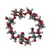  Type: oligosaccharide, Oligosaccharide / Class: Drug delivery / Mass: 1153.001 Da / Num. of mol.: 1 Type: oligosaccharide, Oligosaccharide / Class: Drug delivery / Mass: 1153.001 Da / Num. of mol.: 1Source method: isolated from a genetically manipulated source Details: cyclic oligosaccharide / References: beta-cyclodextrin | ||||||
| #3: Chemical | ChemComp-GOL /   Mass: 92.094 Da / Num. of mol.: 5 / Source method: obtained synthetically / Formula: C3H8O3 Mass: 92.094 Da / Num. of mol.: 5 / Source method: obtained synthetically / Formula: C3H8O3#4: Water | ChemComp-HOH / |  Mass: 18.015 Da / Num. of mol.: 226 / Source method: isolated from a natural source / Formula: H2O Mass: 18.015 Da / Num. of mol.: 226 / Source method: isolated from a natural source / Formula: H2OHas ligand of interest | Y | Has protein modification | N | |
-Experimental details
-Experiment
| Experiment | Method: X-RAY DIFFRACTION / Number of used crystals: 1 |
|---|
- Sample preparation
| Crystal | Density Matthews: 2.26 Å3/Da / Density % sol: 45.61 % |
|---|---|
| Crystal grow | Temperature: 338 K / Method: vapor diffusion, sitting drop / Details: 0.1M Sodium citrate, pH 4.2, 20% PEG 3350 |
-Data collection
| Diffraction | Mean temperature: 100 K / Serial crystal experiment: N |
|---|---|
| Diffraction source | Source: SYNCHROTRON / Site: SSRF  / Beamline: BL02U1 / Wavelength: 0.97918 Å / Beamline: BL02U1 / Wavelength: 0.97918 Å |
| Detector | Type: DECTRIS EIGER2 S 9M / Detector: PIXEL / Date: May 1, 2024 |
| Radiation | Protocol: SINGLE WAVELENGTH / Monochromatic (M) / Laue (L): M / Scattering type: x-ray |
| Radiation wavelength | Wavelength: 0.97918 Å / Relative weight: 1 |
| Reflection | Resolution: 1.88→33.56 Å / Num. obs: 42138 / % possible obs: 99.8 % / Redundancy: 6.8 % / CC1/2: 0.996 / Net I/σ(I): 9 |
| Reflection shell | Resolution: 1.88→1.93 Å / Redundancy: 6.9 % / Rmerge(I) obs: 1.381 / Num. unique obs: 3111 / CC1/2: 0.474 / Rpim(I) all: 0.562 / Rrim(I) all: 1.493 |
- Processing
| Software |
| |||||||||||||||||||||||||||||||||||||||||||||||||||||||||||||||||||||||||||||||||||||||||||||||||||||||||||||||||||||||||||||
|---|---|---|---|---|---|---|---|---|---|---|---|---|---|---|---|---|---|---|---|---|---|---|---|---|---|---|---|---|---|---|---|---|---|---|---|---|---|---|---|---|---|---|---|---|---|---|---|---|---|---|---|---|---|---|---|---|---|---|---|---|---|---|---|---|---|---|---|---|---|---|---|---|---|---|---|---|---|---|---|---|---|---|---|---|---|---|---|---|---|---|---|---|---|---|---|---|---|---|---|---|---|---|---|---|---|---|---|---|---|---|---|---|---|---|---|---|---|---|---|---|---|---|---|---|---|---|
| Refinement | Method to determine structure: MOLECULAR REPLACEMENT / Resolution: 1.88→33.558 Å / SU ML: 0.21 / Cross valid method: FREE R-VALUE / σ(F): 1.34 / Phase error: 19.36 / Stereochemistry target values: ML
| |||||||||||||||||||||||||||||||||||||||||||||||||||||||||||||||||||||||||||||||||||||||||||||||||||||||||||||||||||||||||||||
| Solvent computation | Shrinkage radii: 0.9 Å / VDW probe radii: 1.11 Å / Solvent model: FLAT BULK SOLVENT MODEL | |||||||||||||||||||||||||||||||||||||||||||||||||||||||||||||||||||||||||||||||||||||||||||||||||||||||||||||||||||||||||||||
| Refinement step | Cycle: LAST / Resolution: 1.88→33.558 Å
| |||||||||||||||||||||||||||||||||||||||||||||||||||||||||||||||||||||||||||||||||||||||||||||||||||||||||||||||||||||||||||||
| Refine LS restraints |
| |||||||||||||||||||||||||||||||||||||||||||||||||||||||||||||||||||||||||||||||||||||||||||||||||||||||||||||||||||||||||||||
| LS refinement shell |
| |||||||||||||||||||||||||||||||||||||||||||||||||||||||||||||||||||||||||||||||||||||||||||||||||||||||||||||||||||||||||||||
| Refinement TLS params. | Method: refined / Refine-ID: X-RAY DIFFRACTION
| |||||||||||||||||||||||||||||||||||||||||||||||||||||||||||||||||||||||||||||||||||||||||||||||||||||||||||||||||||||||||||||
| Refinement TLS group |
|
