 Movie
Movie Controller

[English] 日本語
 Yorodumi
- PDB-9j7h: Crystal structure of 3-deoxy-D-arabino-heptulosonate-7-phosphate ... -
+ Open data

- Basic information
| Entry | Database: PDB / ID: 9j7h | ||||||
|---|---|---|---|---|---|---|---|
| Title | Crystal structure of 3-deoxy-D-arabino-heptulosonate-7-phosphate synthase (DAHP synthase) from Providencia alcalifaciens complexed with quinic acid | ||||||
 Components Components | Phospho-2-dehydro-3-deoxyheptonate aldolase | ||||||
 Keywords Keywords | TRANSFERASE / DAHP Synthase / barrel domain / inhibitor / quinic acid | ||||||
| Function / homology |  Function and homology information Function and homology information3-deoxy-7-phosphoheptulonate synthase / 3-deoxy-7-phosphoheptulonate synthase activity / chorismate biosynthetic process / aromatic amino acid biosynthetic process / amino acid biosynthetic process / identical protein binding / cytoplasm Similarity search - Function | ||||||
| Biological species |  Providencia alcalifaciens (bacteria) Providencia alcalifaciens (bacteria) | ||||||
| Method |  X-RAY DIFFRACTION / MOLECULAR REPLACEMENT / Resolution: 2.68 Å X-RAY DIFFRACTION / MOLECULAR REPLACEMENT / Resolution: 2.68 Å | ||||||
 Authors Authors | Jangid, K. / Mahto, J.K. / Kumar, K.A. / Kumar, P. | ||||||
| Funding support |  India, 1items India, 1items
| ||||||
 Citation Citation | Journal: Arch.Biochem.Biophys. / Year: 2025 Title: Structural and biochemical analyses reveal quinic acid inhibits DAHP synthase a key player in shikimate pathway. Authors: Jangid, K. / Mahto, J.K. / Kumar, K.A. / Dhaka, P. / Sharma, A. / Tariq, A. / Sharma, A.K. / Kumar, P. | ||||||
| History |
|
- Structure visualization
| Structure viewer | Molecule: MolmilJmol/JSmol |
|---|
- Downloads & links
-Download
| PDBx/mmCIF format | 9j7h.cif.gz | 453.3 KB | Display | PDBx/mmCIF format |
|---|---|---|---|---|
| PDB format | pdb9j7h.ent.gz | 373.6 KB | Display | PDB format |
| PDBx/mmJSON format | 9j7h.json.gz | Tree view | PDBx/mmJSON format | |
| Others |  Other downloads Other downloads |
-Validation report
| Arichive directory | https://data.pdbj.org/pub/pdb/validation_reports/j7/9j7hftp://data.pdbj.org/pub/pdb/validation_reports/j7/9j7h | HTTPS FTP |
|---|
-Related structure data
| Related structure data |  9j7sC 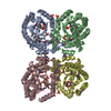 1kflS S: Starting model for refinement C: citing same article ( |
|---|---|
| Similar structure data |
-Links
 PDBj
PDBj- Assembly
| Deposited unit | 
| ||||||||
|---|---|---|---|---|---|---|---|---|---|
| 1 |
| ||||||||
| Unit cell |
|
-Components
| #1: Protein | Mass: 38761.996 Da / Num. of mol.: 4 Source method: isolated from a genetically manipulated source Source: (gene. exp.) Providencia alcalifaciens (bacteria) / Gene: PROVALCAL_03276 / Production host: References: UniProt: B6XIT1, 3-deoxy-7-phosphoheptulonate synthase #2: Chemical | ChemComp-QIC / (   Mass: 192.167 Da / Num. of mol.: 4 / Source method: obtained synthetically / Formula: C7H12O6 / Feature type: SUBJECT OF INVESTIGATION Mass: 192.167 Da / Num. of mol.: 4 / Source method: obtained synthetically / Formula: C7H12O6 / Feature type: SUBJECT OF INVESTIGATION#3: Water | ChemComp-HOH / |  Mass: 18.015 Da / Num. of mol.: 22 / Source method: isolated from a natural source / Formula: H2O Mass: 18.015 Da / Num. of mol.: 22 / Source method: isolated from a natural source / Formula: H2OHas ligand of interest | Y | Has protein modification | Y | |
|---|
-Experimental details
-Experiment
| Experiment | Method: X-RAY DIFFRACTION / Number of used crystals: 1 |
|---|
- Sample preparation
| Crystal | Density Matthews: 2.59 Å3/Da / Density % sol: 52.53 % |
|---|---|
| Crystal grow | Temperature: 293 K / Method: vapor diffusion, sitting drop Details: 25%w/v PEG3350, 0.2M Magnesium chloride, 0.1M bis Tris PH-5.5, 20% ETG |
-Data collection
| Diffraction | Mean temperature: 100 K / Serial crystal experiment: N |
|---|---|
| Diffraction source | Source: ROTATING ANODE / Type: RIGAKU MICROMAX-007 HF / Wavelength: 1.54184 Å |
| Detector | Type: RIGAKU HyPix-6000HE / Detector: PIXEL / Date: Nov 15, 2022 |
| Radiation | Protocol: SINGLE WAVELENGTH / Monochromatic (M) / Laue (L): M / Scattering type: x-ray |
| Radiation wavelength | Wavelength: 1.54184 Å / Relative weight: 1 |
| Reflection | Resolution: 2.68→25.21 Å / Num. obs: 43906 / % possible obs: 96.8 % / Redundancy: 3.5 % / CC1/2: 0.943 / Net I/σ(I): 4.5 |
| Reflection shell | Resolution: 2.68→2.78 Å / Mean I/σ(I) obs: 1.2 / Num. unique obs: 4373 / CC1/2: 0.699 / % possible all: 97.8 |
- Processing
| Software |
| ||||||||||||||||||||||||||||||||||||||||||||||||||||||||||||||||||||||||||||||||||||||||||||||||||||||||||||||||||||||||||||||||||||||||||||||||||||||||||||||||||||||||||||||||||||||
|---|---|---|---|---|---|---|---|---|---|---|---|---|---|---|---|---|---|---|---|---|---|---|---|---|---|---|---|---|---|---|---|---|---|---|---|---|---|---|---|---|---|---|---|---|---|---|---|---|---|---|---|---|---|---|---|---|---|---|---|---|---|---|---|---|---|---|---|---|---|---|---|---|---|---|---|---|---|---|---|---|---|---|---|---|---|---|---|---|---|---|---|---|---|---|---|---|---|---|---|---|---|---|---|---|---|---|---|---|---|---|---|---|---|---|---|---|---|---|---|---|---|---|---|---|---|---|---|---|---|---|---|---|---|---|---|---|---|---|---|---|---|---|---|---|---|---|---|---|---|---|---|---|---|---|---|---|---|---|---|---|---|---|---|---|---|---|---|---|---|---|---|---|---|---|---|---|---|---|---|---|---|---|---|
| Refinement | Method to determine structure: MOLECULAR REPLACEMENT Starting model: 1KFL Resolution: 2.68→25.21 Å / Cor.coef. Fo:Fc: 0.845 / Cor.coef. Fo:Fc free: 0.787 / SU B: 32.288 / SU ML: 0.301 / Cross valid method: THROUGHOUT / ESU R: 1.005 / ESU R Free: 0.396 / Stereochemistry target values: MAXIMUM LIKELIHOOD / Details: HYDROGENS HAVE BEEN ADDED IN THE RIDING POSITIONS
| ||||||||||||||||||||||||||||||||||||||||||||||||||||||||||||||||||||||||||||||||||||||||||||||||||||||||||||||||||||||||||||||||||||||||||||||||||||||||||||||||||||||||||||||||||||||
| Solvent computation | Ion probe radii: 0.8 Å / Shrinkage radii: 0.8 Å / VDW probe radii: 1.2 Å / Solvent model: MASK | ||||||||||||||||||||||||||||||||||||||||||||||||||||||||||||||||||||||||||||||||||||||||||||||||||||||||||||||||||||||||||||||||||||||||||||||||||||||||||||||||||||||||||||||||||||||
| Displacement parameters | Biso mean: 34.944 Å2
| ||||||||||||||||||||||||||||||||||||||||||||||||||||||||||||||||||||||||||||||||||||||||||||||||||||||||||||||||||||||||||||||||||||||||||||||||||||||||||||||||||||||||||||||||||||||
| Refinement step | Cycle: 1 / Resolution: 2.68→25.21 Å
| ||||||||||||||||||||||||||||||||||||||||||||||||||||||||||||||||||||||||||||||||||||||||||||||||||||||||||||||||||||||||||||||||||||||||||||||||||||||||||||||||||||||||||||||||||||||
| Refine LS restraints |
|