 Movie
Movie Controller


+ Open data

- Basic information
| Entry | Database: PDB / ID: 9izf | |||||||||||||||||||||||||||||||||||||||
|---|---|---|---|---|---|---|---|---|---|---|---|---|---|---|---|---|---|---|---|---|---|---|---|---|---|---|---|---|---|---|---|---|---|---|---|---|---|---|---|---|
| Title | Cryo-EM structure of LPA1-Gi complex with LPA | |||||||||||||||||||||||||||||||||||||||
 Components Components |
| |||||||||||||||||||||||||||||||||||||||
 Keywords Keywords | MEMBRANE PROTEIN / GPCR / SIGNALING PROTEIN | |||||||||||||||||||||||||||||||||||||||
| Function / homology |  Function and homology information Function and homology informationcellular response to 1-oleoyl-sn-glycerol 3-phosphate / lysophosphatidic acid receptor activity / G beta:gamma signalling through PLC beta / Presynaptic function of Kainate receptors / Prostacyclin signalling through prostacyclin receptor / G alpha (z) signalling events / Glucagon-type ligand receptors / G beta:gamma signalling through PI3Kgamma / G beta:gamma signalling through CDC42 / positive regulation of smooth muscle cell chemotaxis ...cellular response to 1-oleoyl-sn-glycerol 3-phosphate / lysophosphatidic acid receptor activity / G beta:gamma signalling through PLC beta / Presynaptic function of Kainate receptors / Prostacyclin signalling through prostacyclin receptor / G alpha (z) signalling events / Glucagon-type ligand receptors / G beta:gamma signalling through PI3Kgamma / G beta:gamma signalling through CDC42 / positive regulation of smooth muscle cell chemotaxis / Adrenaline,noradrenaline inhibits insulin secretion / ADP signalling through P2Y purinoceptor 12 / Cooperation of PDCL (PhLP1) and TRiC/CCT in G-protein beta folding / Thromboxane signalling through TP receptor / G beta:gamma signalling through BTK / Thrombin signalling through proteinase activated receptors (PARs) / Activation of G protein gated Potassium channels / Inhibition of voltage gated Ca2+ channels via Gbeta/gamma subunits / lysophosphatidic acid binding / G alpha (s) signalling events / Lysosphingolipid and LPA receptors / Ca2+ pathway / G-protein activation / Extra-nuclear estrogen signaling / G alpha (12/13) signalling events / G alpha (q) signalling events / Vasopressin regulates renal water homeostasis via Aquaporins / negative regulation of cilium assembly / GPER1 signaling / Glucagon-like Peptide-1 (GLP1) regulates insulin secretion / corpus callosum development / ADP signalling through P2Y purinoceptor 1 / High laminar flow shear stress activates signaling by PIEZO1 and PECAM1:CDH5:KDR in endothelial cells / G alpha (i) signalling events / oligodendrocyte development / cellular response to oxygen levels / optic nerve development / regulation of synaptic vesicle cycle / regulation of postsynaptic neurotransmitter receptor internalization / positive regulation of dendritic spine development / positive regulation of Rho protein signal transduction / G-protein alpha-subunit binding / negative regulation of cAMP/PKA signal transduction / positive regulation of stress fiber assembly / regulation of eating behavior / adenylate cyclase inhibitor activity / myelination / positive regulation of protein localization to cell cortex / T cell migration / positive regulation of relaxation of smooth muscle / Adenylate cyclase inhibitory pathway / neurogenesis / D2 dopamine receptor binding / adenylate cyclase-inhibiting serotonin receptor signaling pathway / cerebellum development / G protein-coupled serotonin receptor binding / cellular response to forskolin / mast cell degranulation / regulation of mitotic spindle organization / dendritic shaft / chemokine-mediated signaling pathway / cell chemotaxis / PDZ domain binding / Regulation of insulin secretion / neuropeptide signaling pathway / response to prostaglandin E / electron transport chain / positive regulation of cholesterol biosynthetic process / G protein-coupled receptor binding / response to peptide hormone / GABA-ergic synapse / G protein-coupled receptor activity / G-protein beta/gamma-subunit complex binding / adenylate cyclase-modulating G protein-coupled receptor signaling pathway / adenylate cyclase-inhibiting G protein-coupled receptor signaling pathway / Olfactory Signaling Pathway / Activation of the phototransduction cascade / G protein-coupled acetylcholine receptor signaling pathway / G beta:gamma signalling through PLC beta / Presynaptic function of Kainate receptors / Thromboxane signalling through TP receptor / Activation of G protein gated Potassium channels / Inhibition of voltage gated Ca2+ channels via Gbeta/gamma subunits / G-protein activation / Glucagon signaling in metabolic regulation / G beta:gamma signalling through CDC42 / Prostacyclin signalling through prostacyclin receptor / Synthesis, secretion, and inactivation of Glucagon-like Peptide-1 (GLP-1) / G beta:gamma signalling through BTK / photoreceptor disc membrane / GDP binding / ADP signalling through P2Y purinoceptor 12 / Glucagon-type ligand receptors / Sensory perception of sweet, bitter, and umami (glutamate) taste / Adrenaline,noradrenaline inhibits insulin secretion / Vasopressin regulates renal water homeostasis via Aquaporins / regulation of cell shape / Glucagon-like Peptide-1 (GLP1) regulates insulin secretion / G alpha (z) signalling events / negative regulation of neuron projection development Similarity search - Function | |||||||||||||||||||||||||||||||||||||||
| Biological species |   Homo sapiens (human) Homo sapiens (human)synthetic construct (others)  | |||||||||||||||||||||||||||||||||||||||
| Method | ELECTRON MICROSCOPY / single particle reconstruction / cryo EM / Resolution: 3.14 Å | |||||||||||||||||||||||||||||||||||||||
 Authors Authors | Suzuki, S. / Nishikawa, K. / Kamegawa, A. / Hiroaki, Y. / Suzuki, H. / Fujiyoshi, Y. | |||||||||||||||||||||||||||||||||||||||
| Funding support |  Japan, 1items Japan, 1items
| |||||||||||||||||||||||||||||||||||||||
 Citation Citation | Journal: J Struct Biol / Year: 2025 Title: Structural insights into the engagement of lysophosphatidic acid receptor 1 with different G proteins. Authors: Shota Suzuki / Kotaro Tanaka / Akiko Kamegawa / Kouki Nishikawa / Hiroshi Suzuki / Atsunori Oshima / Yoshinori Fujiyoshi / Abstract: Lysophosphatidic acid (LPA) and sphingosine-1-phosphate (S1P) are bioactive lysophospholipids derived from cell membranes that activate the endothelial differentiation gene family of G protein- ...Lysophosphatidic acid (LPA) and sphingosine-1-phosphate (S1P) are bioactive lysophospholipids derived from cell membranes that activate the endothelial differentiation gene family of G protein-coupled receptors. Activation of these receptors triggers multiple downstream signaling cascades through G proteins such as Gi/o, Gq/11, and G12/13. Therefore, LPA and S1P mediate several physiological processes, including cytoskeletal dynamics, neurite retraction, cell migration, cell proliferation, and intracellular ion fluxes. The basis for the G-protein coupling selectivity of EDG receptors, however, remains unknown. Here, we present cryo-electron microscopy structures of LPA-activated LPA1 in complexes with G, G, and G heterotrimers Comparison of the three LPA1-G protein structures shows clearly different conformations of intracellular loop 2 (ICL2) and ICL3 that are likely induced by the different Gα protein interfaces. Interestingly, this G-protein interface interaction is a common feature of LPA and S1P receptors. Our findings provide clues to understanding the promiscuity of G-protein coupling in the endothelial differentiation gene family. | |||||||||||||||||||||||||||||||||||||||
| History |
|
- Structure visualization
| Structure viewer | Molecule: MolmilJmol/JSmol |
|---|
- Downloads & links
-Download
| PDBx/mmCIF format | 9izf.cif.gz | 276 KB | Display | PDBx/mmCIF format |
|---|---|---|---|---|
| PDB format | pdb9izf.ent.gz | Display | PDB format | |
| PDBx/mmJSON format | 9izf.json.gz | Tree view | PDBx/mmJSON format | |
| Others |  Other downloads Other downloads |
-Validation report
| Arichive directory | https://data.pdbj.org/pub/pdb/validation_reports/iz/9izfftp://data.pdbj.org/pub/pdb/validation_reports/iz/9izf | HTTPS FTP |
|---|
-Related structure data
| Related structure data |  61031MC 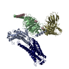 9izgC  9izhC  61034 61035 61036 C: citing same article ( M: map data used to model this data |
|---|---|
| Similar structure data |


-Links
 PDBj
PDBj


- Assembly
| Deposited unit | 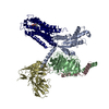
|
|---|---|
| 1 |
|
-Components
-Guanine nucleotide-binding protein ... , 3 types, 3 molecules ABC
| #2: Protein | Mass: 40446.047 Da / Num. of mol.: 1 Source method: isolated from a genetically manipulated source Source: (gene. exp.) Homo sapiens (human) / Gene: GNAI1 / Production host:   Spodoptera frugiperda (fall armyworm) / References: UniProt: P63096 Spodoptera frugiperda (fall armyworm) / References: UniProt: P63096 |
|---|---|
| #3: Protein | Mass: 41772.562 Da / Num. of mol.: 1 Source method: isolated from a genetically manipulated source Details: N-terminal His tag C-terminal GS linker and HiBiT tag Source: (gene. exp.) Homo sapiens (human) / Gene: GNB1 / Production host: Spodoptera frugiperda (fall armyworm) / References: UniProt: P62873 |
| #4: Protein | Mass: 7729.947 Da / Num. of mol.: 1 Source method: isolated from a genetically manipulated source Source: (gene. exp.) Spodoptera frugiperda (fall armyworm) / References: UniProt: P63213 |
-Protein / Antibody / Non-polymers , 3 types, 3 molecules RS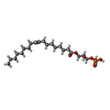
| #1: Protein | Mass: 73188.500 Da / Num. of mol.: 1 Source method: isolated from a genetically manipulated source Details: N-terminal HA signal sequence, FLAG tag C-terminal LgBiT Source: (gene. exp.) Homo sapiens (human), (gene. exp.) synthetic construct (others)Gene: cybC, LPAR1, EDG2, LPA1 / Production host: Spodoptera frugiperda (fall armyworm) / References: UniProt: P0ABE7, UniProt: Q92633 |
|---|---|
| #5: Antibody | Mass: 26466.486 Da / Num. of mol.: 1 Source method: isolated from a genetically manipulated source Source: (gene. exp.) synthetic construct (others) / Production host: Spodoptera frugiperda (fall armyworm) |
| #6: Chemical | ChemComp-NKP / ( Mass: 436.520 Da / Num. of mol.: 1 / Source method: obtained synthetically / Formula: C21H41O7P / Feature type: SUBJECT OF INVESTIGATION Mass: 436.520 Da / Num. of mol.: 1 / Source method: obtained synthetically / Formula: C21H41O7P / Feature type: SUBJECT OF INVESTIGATION |
-Details
| Has ligand of interest | Y |
|---|---|
| Has protein modification | Y |
-Experimental details
-Experiment
| Experiment | Method: ELECTRON MICROSCOPY |
|---|---|
| EM experiment | Aggregation state: PARTICLE / 3D reconstruction method: single particle reconstruction |
- Sample preparation
| Component | Name: Multiprotein complex / Type: COMPLEX / Entity ID: #1-#5 / Source: RECOMBINANT |
|---|---|
| Molecular weight | Experimental value: NO |
| Source (natural) | Organism: Homo sapiens (human) |
| Source (recombinant) | Organism: Spodoptera frugiperda (fall armyworm) |
| Buffer solution | pH: 7.4 |
| Specimen | Conc.: 15 mg/ml / Embedding applied: NO / Shadowing applied: NO / Staining applied: NO / Vitrification applied: YES / Details: This sample was monodisperse |
| Vitrification | Cryogen name: ETHANE / Humidity: 100 % / Chamber temperature: 298 K |
- Electron microscopy imaging
| Microscopy | Model: JEOL CRYO ARM 300 |
|---|---|
| Electron gun | Electron source:  FIELD EMISSION GUN / Accelerating voltage: 300 kV / Illumination mode: FLOOD BEAM FIELD EMISSION GUN / Accelerating voltage: 300 kV / Illumination mode: FLOOD BEAM |
| Electron lens | Mode: BRIGHT FIELD / Nominal defocus max: 2000 nm / Nominal defocus min: 1000 nm |
| Image recording | Electron dose: 71 e/Å2 / Detector mode: COUNTING / Film or detector model: GATAN K2 SUMMIT (4k x 4k) |
- Processing
| EM software | Name: PHENIX / Category: model refinement | ||||||||||||||||||||||||
|---|---|---|---|---|---|---|---|---|---|---|---|---|---|---|---|---|---|---|---|---|---|---|---|---|---|
| CTF correction | Type: PHASE FLIPPING AND AMPLITUDE CORRECTION | ||||||||||||||||||||||||
| 3D reconstruction | Resolution: 3.14 Å / Resolution method: FSC 0.143 CUT-OFF / Num. of particles: 178606 / Symmetry type: POINT | ||||||||||||||||||||||||
| Refine LS restraints |
|
