+ Open data
- Basic information
| Entry | Database: PDB / ID: 9it2 | ||||||||||||||||||||||||
|---|---|---|---|---|---|---|---|---|---|---|---|---|---|---|---|---|---|---|---|---|---|---|---|---|---|
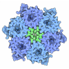
| Title | Cryo-EM structure of urease from Ureaplasma parvum | ||||||||||||||||||||||||
 Components Components | (Urease subunit ...) x 3 | ||||||||||||||||||||||||
 Keywords Keywords | HYDROLASE / urease | ||||||||||||||||||||||||
| Function / homology |  Function and homology information Function and homology informationurease complex / urease / urease activity / urea catabolic process / nickel cation binding / cytoplasm Similarity search - Function | ||||||||||||||||||||||||
| Biological species | Ureaplasma parvum serovar 3 | ||||||||||||||||||||||||
| Method | ELECTRON MICROSCOPY / single particle reconstruction / cryo EM / Resolution: 2.03 Å | ||||||||||||||||||||||||
 Authors Authors | Fujita, J. / Namba, K. / Wu, H.N. / Yanagihara, I. | ||||||||||||||||||||||||
| Funding support |  Japan, 7items Japan, 7items
| ||||||||||||||||||||||||
 Citation Citation |  Journal: J Mol Biol / Year: 2025 Journal: J Mol Biol / Year: 2025Title: Structural Analysis and Molecular Dynamics Simulations of Urease From Ureaplasma parvum. Authors: Heng Ning Wu / Junso Fujita / Yukiko Nakura / Masao Inoue / Koichiro Suzuki / Toru Ekimoto / Bingjie Yin / Yohta Fukuda / Kazuo Harada / Tsuyoshi Inoue / Mitsunori Ikeguchi / Keiichi Namba / Itaru Yanagihara /  Abstract: Ureaplasma is one of the smallest pathogenic bacteria, generating approximately 95% of its adenosine triphosphate (ATP) solely through urease. Studies on Ureaplasma parvum, a species of Ureaplasma, ...Ureaplasma is one of the smallest pathogenic bacteria, generating approximately 95% of its adenosine triphosphate (ATP) solely through urease. Studies on Ureaplasma parvum, a species of Ureaplasma, have confirmed that adding urease inhibitors inhibits bacterial growth. The K and V of the urease-mediated reaction were estimated to be 4.3 ± 0.2 mM and 3,333.3 ± 38.0 μmol NH/min/mg protein, respectively. The cryo-electron microscopy (cryo-EM) structure of Ureaplasma parvum urease (UPU) at a resolution of 2.03 Å reveals a trimer of heterotrimers comprising three proteins: UreA, UreB, and UreC. The active site is well conserved among the known ureases. However, the V of UPU was higher than that of most known ureases, including those ureases derived from Sporosarcina pasteurii (SPU) and Klebsiella aerogenes (KAU) with identical oligomeric state. All-atom molecular dynamics simulations showed that the flap and UreB are more open in UPU than SPU and KAU. His-tagged wild-type recombinant UPU (WT-rUPU) revealed estimated K and V values of 4.1 ± 0.3 mM and 769.2 ± 7.4 µmol NH/min/mg protein, respectively. Amino acid substitutions of recombinant UPUs within the flap region to SPU. Amongst the flap region variants, the V of K331N variant was 48-fold lower than that of WT-rUPU. ICP-MS analysis reveals that one molecule of UPU, WT-rUPU, and K331N-rUPU contains 3.7, 0.8, and 0.1 Ni atoms, respectively, suggesting that a wide-open flap of urease may contribute to delivering nickel into the enzyme, resulting in a high V. Ureaplasma evolved highly efficient UPU through a few amino acid substitutions in the disorganized loop of the mobile flap region. | ||||||||||||||||||||||||
| History |
|
- Structure visualization
| Structure viewer | Molecule:  Molmil Molmil Jmol/JSmol Jmol/JSmol |
|---|
- Downloads & links
- Download
| PDBx/mmCIF format |  9it2.cif.gz 9it2.cif.gz | 416.1 KB | Display |  PDBx/mmCIF format PDBx/mmCIF format |
|---|---|---|---|---|
| PDB format |  pdb9it2.ent.gz pdb9it2.ent.gz | 342 KB | Display |  PDB format PDB format |
| PDBx/mmJSON format |  9it2.json.gz 9it2.json.gz | Tree view |  PDBx/mmJSON format PDBx/mmJSON format | |
| Others |  Other downloads Other downloads |
-Validation report
| Arichive directory |  https://data.pdbj.org/pub/pdb/validation_reports/it/9it2 https://data.pdbj.org/pub/pdb/validation_reports/it/9it2 ftp://data.pdbj.org/pub/pdb/validation_reports/it/9it2 ftp://data.pdbj.org/pub/pdb/validation_reports/it/9it2 | HTTPS FTP |
|---|
-Related structure data
| Related structure data |  60854MC M: map data used to model this data C: citing same article ( |
|---|---|
| Similar structure data | Similarity search - Function & homology  F&H Search F&H Search |
- Links
Links
- Assembly
| Deposited unit | 
|
|---|---|
| 1 |
|
- Components
-Urease subunit ... , 3 types, 9 molecules ADGBEHCFI
| #1: Protein | Mass: 11234.106 Da / Num. of mol.: 3 / Source method: isolated from a natural source Source: (natural)  Ureaplasma parvum serovar 3 (strain ATCC 700970) (bacteria) Ureaplasma parvum serovar 3 (strain ATCC 700970) (bacteria)References: UniProt: P0C7K9, urease #2: Protein | Mass: 13529.323 Da / Num. of mol.: 3 / Source method: isolated from a natural source Source: (natural)  Ureaplasma parvum serovar 3 (strain ATCC 700970) (bacteria) Ureaplasma parvum serovar 3 (strain ATCC 700970) (bacteria)References: UniProt: P0C7K8, urease #3: Protein | Mass: 64661.324 Da / Num. of mol.: 3 / Source method: isolated from a natural source Source: (natural)  Ureaplasma parvum serovar 3 (strain ATCC 700970) (bacteria) Ureaplasma parvum serovar 3 (strain ATCC 700970) (bacteria)References: UniProt: P0C7K7, urease |
|---|
-Non-polymers , 3 types, 443 molecules 


| #4: Chemical | | #5: Chemical | ChemComp-NI / #6: Water | ChemComp-HOH / | |
|---|
-Details
| Has ligand of interest | Y |
|---|---|
| Has protein modification | Y |
-Experimental details
-Experiment
| Experiment | Method: ELECTRON MICROSCOPY |
|---|---|
| EM experiment | Aggregation state: PARTICLE / 3D reconstruction method: single particle reconstruction |
- Sample preparation
| Component | Name: Urease / Type: COMPLEX / Entity ID: #1-#3 / Source: NATURAL | ||||||||||||||||||||
|---|---|---|---|---|---|---|---|---|---|---|---|---|---|---|---|---|---|---|---|---|---|
| Molecular weight | Experimental value: NO | ||||||||||||||||||||
| Source (natural) | Organism:  Ureaplasma parvum serovar 3 (strain ATCC 700970) (bacteria) Ureaplasma parvum serovar 3 (strain ATCC 700970) (bacteria) | ||||||||||||||||||||
| Buffer solution | pH: 8 | ||||||||||||||||||||
| Buffer component |
| ||||||||||||||||||||
| Specimen | Conc.: 0.78 mg/ml / Embedding applied: NO / Shadowing applied: NO / Staining applied: NO / Vitrification applied: YES | ||||||||||||||||||||
| Specimen support | Grid material: COPPER / Grid mesh size: 200 divisions/in. / Grid type: Quantifoil R1.2/1.3 | ||||||||||||||||||||
| Vitrification | Instrument: FEI VITROBOT MARK IV / Cryogen name: ETHANE / Humidity: 100 % / Chamber temperature: 281 K |
- Electron microscopy imaging
| Microscopy | Model: JEOL CRYO ARM 300 |
|---|---|
| Electron gun | Electron source:  FIELD EMISSION GUN / Accelerating voltage: 300 kV / Illumination mode: FLOOD BEAM FIELD EMISSION GUN / Accelerating voltage: 300 kV / Illumination mode: FLOOD BEAM |
| Electron lens | Mode: BRIGHT FIELD / Nominal magnification: 60000 X / Nominal defocus max: 2000 nm / Nominal defocus min: 500 nm / Cs: 2.7 mm |
| Specimen holder | Cryogen: NITROGEN / Specimen holder model: JEOL CRYOSPECPORTER |
| Image recording | Average exposure time: 1.8 sec. / Electron dose: 40 e/Å2 / Film or detector model: GATAN K3 (6k x 4k) / Num. of grids imaged: 1 |
| EM imaging optics | Energyfilter name: In-column Omega Filter / Energyfilter slit width: 20 eV |
- Processing
| EM software |
| ||||||||||||||||||||||||||||||||||||
|---|---|---|---|---|---|---|---|---|---|---|---|---|---|---|---|---|---|---|---|---|---|---|---|---|---|---|---|---|---|---|---|---|---|---|---|---|---|
| CTF correction | Type: PHASE FLIPPING AND AMPLITUDE CORRECTION | ||||||||||||||||||||||||||||||||||||
| Particle selection | Num. of particles selected: 3333272 | ||||||||||||||||||||||||||||||||||||
| Symmetry | Point symmetry: C3 (3 fold cyclic) | ||||||||||||||||||||||||||||||||||||
| 3D reconstruction | Resolution: 2.03 Å / Resolution method: FSC 0.143 CUT-OFF / Num. of particles: 207602 / Algorithm: FOURIER SPACE / Symmetry type: POINT | ||||||||||||||||||||||||||||||||||||
| Atomic model building | Space: REAL | ||||||||||||||||||||||||||||||||||||
| Atomic model building | Source name: AlphaFold / Type: in silico model | ||||||||||||||||||||||||||||||||||||
| Refine LS restraints |
|
 Movie
Movie Controller


 PDBj
PDBj


