+ Open data
- Basic information
| Entry |  | ||||||||||||||||||||||||
|---|---|---|---|---|---|---|---|---|---|---|---|---|---|---|---|---|---|---|---|---|---|---|---|---|---|
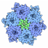
| Title | Cryo-EM structure of urease from Ureaplasma parvum | ||||||||||||||||||||||||
 Map data Map data | |||||||||||||||||||||||||
 Sample Sample |
| ||||||||||||||||||||||||
 Keywords Keywords | urease / HYDROLASE | ||||||||||||||||||||||||
| Function / homology |  Function and homology information Function and homology informationurease complex / urease / urease activity / urea catabolic process / nickel cation binding / cytoplasm Similarity search - Function | ||||||||||||||||||||||||
| Biological species |  Ureaplasma parvum serovar 3 (strain ATCC 700970) (bacteria) Ureaplasma parvum serovar 3 (strain ATCC 700970) (bacteria) | ||||||||||||||||||||||||
| Method | single particle reconstruction / cryo EM / Resolution: 2.03 Å | ||||||||||||||||||||||||
 Authors Authors | Fujita J / Namba K / Wu HN / Yanagihara I | ||||||||||||||||||||||||
| Funding support |  Japan, 7 items Japan, 7 items
| ||||||||||||||||||||||||
 Citation Citation |  Journal: J Mol Biol / Year: 2025 Journal: J Mol Biol / Year: 2025Title: Structural Analysis and Molecular Dynamics Simulations of Urease From Ureaplasma parvum. Authors: Heng Ning Wu / Junso Fujita / Yukiko Nakura / Masao Inoue / Koichiro Suzuki / Toru Ekimoto / Bingjie Yin / Yohta Fukuda / Kazuo Harada / Tsuyoshi Inoue / Mitsunori Ikeguchi / Keiichi Namba / Itaru Yanagihara /  Abstract: Ureaplasma is one of the smallest pathogenic bacteria, generating approximately 95% of its adenosine triphosphate (ATP) solely through urease. Studies on Ureaplasma parvum, a species of Ureaplasma, ...Ureaplasma is one of the smallest pathogenic bacteria, generating approximately 95% of its adenosine triphosphate (ATP) solely through urease. Studies on Ureaplasma parvum, a species of Ureaplasma, have confirmed that adding urease inhibitors inhibits bacterial growth. The K and V of the urease-mediated reaction were estimated to be 4.3 ± 0.2 mM and 3,333.3 ± 38.0 μmol NH/min/mg protein, respectively. The cryo-electron microscopy (cryo-EM) structure of Ureaplasma parvum urease (UPU) at a resolution of 2.03 Å reveals a trimer of heterotrimers comprising three proteins: UreA, UreB, and UreC. The active site is well conserved among the known ureases. However, the V of UPU was higher than that of most known ureases, including those ureases derived from Sporosarcina pasteurii (SPU) and Klebsiella aerogenes (KAU) with identical oligomeric state. All-atom molecular dynamics simulations showed that the flap and UreB are more open in UPU than SPU and KAU. His-tagged wild-type recombinant UPU (WT-rUPU) revealed estimated K and V values of 4.1 ± 0.3 mM and 769.2 ± 7.4 µmol NH/min/mg protein, respectively. Amino acid substitutions of recombinant UPUs within the flap region to SPU. Amongst the flap region variants, the V of K331N variant was 48-fold lower than that of WT-rUPU. ICP-MS analysis reveals that one molecule of UPU, WT-rUPU, and K331N-rUPU contains 3.7, 0.8, and 0.1 Ni atoms, respectively, suggesting that a wide-open flap of urease may contribute to delivering nickel into the enzyme, resulting in a high V. Ureaplasma evolved highly efficient UPU through a few amino acid substitutions in the disorganized loop of the mobile flap region. | ||||||||||||||||||||||||
| History |
|
- Structure visualization
| Supplemental images |
|---|
- Downloads & links
-EMDB archive
| Map data |  emd_60854.map.gz emd_60854.map.gz | 168 MB |  EMDB map data format EMDB map data format | |
|---|---|---|---|---|
| Header (meta data) |  emd-60854-v30.xml emd-60854-v30.xml emd-60854.xml emd-60854.xml | 22.9 KB 22.9 KB | Display Display |  EMDB header EMDB header |
| FSC (resolution estimation) |  emd_60854_fsc.xml emd_60854_fsc.xml | 11.8 KB | Display |  FSC data file FSC data file |
| Images |  emd_60854.png emd_60854.png | 70.4 KB | ||
| Masks |  emd_60854_msk_1.map emd_60854_msk_1.map | 178 MB |  Mask map Mask map | |
| Filedesc metadata |  emd-60854.cif.gz emd-60854.cif.gz | 6.8 KB | ||
| Others |  emd_60854_half_map_1.map.gz emd_60854_half_map_1.map.gz emd_60854_half_map_2.map.gz emd_60854_half_map_2.map.gz | 165.4 MB 165.4 MB | ||
| Archive directory |  http://ftp.pdbj.org/pub/emdb/structures/EMD-60854 http://ftp.pdbj.org/pub/emdb/structures/EMD-60854 ftp://ftp.pdbj.org/pub/emdb/structures/EMD-60854 ftp://ftp.pdbj.org/pub/emdb/structures/EMD-60854 | HTTPS FTP |
-Validation report
| Summary document |  emd_60854_validation.pdf.gz emd_60854_validation.pdf.gz | 1006.6 KB | Display |  EMDB validaton report EMDB validaton report |
|---|---|---|---|---|
| Full document |  emd_60854_full_validation.pdf.gz emd_60854_full_validation.pdf.gz | 1006.2 KB | Display | |
| Data in XML |  emd_60854_validation.xml.gz emd_60854_validation.xml.gz | 20.4 KB | Display | |
| Data in CIF |  emd_60854_validation.cif.gz emd_60854_validation.cif.gz | 26.6 KB | Display | |
| Arichive directory |  https://ftp.pdbj.org/pub/emdb/validation_reports/EMD-60854 https://ftp.pdbj.org/pub/emdb/validation_reports/EMD-60854 ftp://ftp.pdbj.org/pub/emdb/validation_reports/EMD-60854 ftp://ftp.pdbj.org/pub/emdb/validation_reports/EMD-60854 | HTTPS FTP |
-Related structure data
| Related structure data |  9it2MC M: atomic model generated by this map C: citing same article ( |
|---|---|
| Similar structure data | Similarity search - Function & homology  F&H Search F&H Search |
- Links
Links
| EMDB pages |  EMDB (EBI/PDBe) / EMDB (EBI/PDBe) /  EMDataResource EMDataResource |
|---|---|
| Related items in Molecule of the Month |
- Map
Map
| File |  Download / File: emd_60854.map.gz / Format: CCP4 / Size: 178 MB / Type: IMAGE STORED AS FLOATING POINT NUMBER (4 BYTES) Download / File: emd_60854.map.gz / Format: CCP4 / Size: 178 MB / Type: IMAGE STORED AS FLOATING POINT NUMBER (4 BYTES) | ||||||||||||||||||||||||||||||||||||
|---|---|---|---|---|---|---|---|---|---|---|---|---|---|---|---|---|---|---|---|---|---|---|---|---|---|---|---|---|---|---|---|---|---|---|---|---|---|
| Projections & slices | Image control
Images are generated by Spider. | ||||||||||||||||||||||||||||||||||||
| Voxel size | X=Y=Z: 0.873 Å | ||||||||||||||||||||||||||||||||||||
| Density |
| ||||||||||||||||||||||||||||||||||||
| Symmetry | Space group: 1 | ||||||||||||||||||||||||||||||||||||
| Details | EMDB XML:
|
-Supplemental data
-Mask #1
| File |  emd_60854_msk_1.map emd_60854_msk_1.map | ||||||||||||
|---|---|---|---|---|---|---|---|---|---|---|---|---|---|
| Projections & Slices |
| ||||||||||||
| Density Histograms |
-Half map: #1
| File | emd_60854_half_map_1.map | ||||||||||||
|---|---|---|---|---|---|---|---|---|---|---|---|---|---|
| Projections & Slices |
| ||||||||||||
| Density Histograms |
-Half map: #2
| File | emd_60854_half_map_2.map | ||||||||||||
|---|---|---|---|---|---|---|---|---|---|---|---|---|---|
| Projections & Slices |
| ||||||||||||
| Density Histograms |
- Sample components
-Entire : Urease
| Entire | Name: Urease |
|---|---|
| Components |
|
-Supramolecule #1: Urease
| Supramolecule | Name: Urease / type: complex / ID: 1 / Parent: 0 / Macromolecule list: #1-#3 |
|---|---|
| Source (natural) | Organism:  Ureaplasma parvum serovar 3 (strain ATCC 700970) (bacteria) Ureaplasma parvum serovar 3 (strain ATCC 700970) (bacteria) |
-Macromolecule #1: Urease subunit gamma
| Macromolecule | Name: Urease subunit gamma / type: protein_or_peptide / ID: 1 / Number of copies: 3 / Enantiomer: LEVO / EC number: urease |
|---|---|
| Source (natural) | Organism:  Ureaplasma parvum serovar 3 (strain ATCC 700970) (bacteria) Ureaplasma parvum serovar 3 (strain ATCC 700970) (bacteria) |
| Molecular weight | Theoretical: 11.234106 KDa |
| Sequence | String: MNLSLREVQK LLITVAADVA RRRLARGLKL NYSEAVALIT DHVMEGARDG KLVADLMQSA REVLRVDQVM EGVDTMVSII QVEVTFPDG TKLVSVHDPI YK UniProtKB: Urease subunit gamma |
-Macromolecule #2: Urease subunit beta
| Macromolecule | Name: Urease subunit beta / type: protein_or_peptide / ID: 2 / Number of copies: 3 / Enantiomer: LEVO / EC number: urease |
|---|---|
| Source (natural) | Organism:  Ureaplasma parvum serovar 3 (strain ATCC 700970) (bacteria) Ureaplasma parvum serovar 3 (strain ATCC 700970) (bacteria) |
| Molecular weight | Theoretical: 13.529323 KDa |
| Sequence | String: MSGSSSQFSP GKLVPGAINF ASGEIVMNEG REAKVISIKN TGDRPIQVGS HFHLFEVNSA LVFFDEKGNE DKERKVAYGR RFDIPSGTA IRFEPGDKKE VSIIDLAGTR EVWGVNGLVN GKLKK UniProtKB: Urease subunit beta |
-Macromolecule #3: Urease subunit alpha
| Macromolecule | Name: Urease subunit alpha / type: protein_or_peptide / ID: 3 / Number of copies: 3 / Enantiomer: LEVO / EC number: urease |
|---|---|
| Source (natural) | Organism:  Ureaplasma parvum serovar 3 (strain ATCC 700970) (bacteria) Ureaplasma parvum serovar 3 (strain ATCC 700970) (bacteria) |
| Molecular weight | Theoretical: 64.661324 KDa |
| Sequence | String: MFKISRKNYS DLYGITTGDS VRLGDTNLWV KVEKDLTTYG EESVFGGGKT LREGMGMNST MKLDDKLGNA EVMDLVITNA LIVDYTGIY KADIGIKNGK IAAIGKSGNP HLTDNVDMIV GISTEISAGE GKIYTAGGLD THVHWLEPEI VPVALDGGIT T VIAGGTGM ...String: MFKISRKNYS DLYGITTGDS VRLGDTNLWV KVEKDLTTYG EESVFGGGKT LREGMGMNST MKLDDKLGNA EVMDLVITNA LIVDYTGIY KADIGIKNGK IAAIGKSGNP HLTDNVDMIV GISTEISAGE GKIYTAGGLD THVHWLEPEI VPVALDGGIT T VIAGGTGM NDGTKATTVS PGKFWVKSAL QAADGLSINA GFLAKGQGME DPIFEQIAAG ACGL(KCX)IHEDW GATGNAID L ALTVADKTDV AVAIHTDTLN EAGFVEHTIA AMKGRTIHAY HTEGAGGGHA PDILETVKYA HILPASTNPT IPYTVNTIA EHLDMLMVCH HLNPKVPEDV AFADSRIRSQ TIAAEDLLHD MGAISIMSSD TLAMGRIGEV ATRTWQMAHK MKAQFGSLKG DSEFSDNNR VKRYISKYTI NPAIAHGVDS YIGSLEVGKL ADIVAWEPKF FGAKPYYVVK MGVIARCVAG DPNASIPTCE P VIMRDQFG TYGRLLTNTS VSFVSKIGLE NGIKEEYKLE KELLPVKNCR SVNKKSMKWN SATPNLEVDP QTFDAAVDFN DL ENWLEQS ASELAKKLKK TSSGKYILDA EPLTEAPLAQ RYFLF UniProtKB: Urease subunit alpha |
-Macromolecule #4: BETA-MERCAPTOETHANOL
| Macromolecule | Name: BETA-MERCAPTOETHANOL / type: ligand / ID: 4 / Number of copies: 3 / Formula: BME |
|---|---|
| Molecular weight | Theoretical: 78.133 Da |
| Chemical component information |  ChemComp-BME: |
-Macromolecule #5: NICKEL (II) ION
| Macromolecule | Name: NICKEL (II) ION / type: ligand / ID: 5 / Number of copies: 6 / Formula: NI |
|---|---|
| Molecular weight | Theoretical: 58.693 Da |
| Chemical component information |  ChemComp-NI: |
-Macromolecule #6: water
| Macromolecule | Name: water / type: ligand / ID: 6 / Number of copies: 434 / Formula: HOH |
|---|---|
| Molecular weight | Theoretical: 18.015 Da |
| Chemical component information |  ChemComp-HOH: |
-Experimental details
-Structure determination
| Method | cryo EM |
|---|---|
 Processing Processing | single particle reconstruction |
| Aggregation state | particle |
- Sample preparation
| Concentration | 0.78 mg/mL | ||||||||||||
|---|---|---|---|---|---|---|---|---|---|---|---|---|---|
| Buffer | pH: 8 Component:
| ||||||||||||
| Grid | Model: Quantifoil R1.2/1.3 / Material: COPPER / Mesh: 200 / Pretreatment - Type: GLOW DISCHARGE | ||||||||||||
| Vitrification | Cryogen name: ETHANE / Chamber humidity: 100 % / Chamber temperature: 281 K / Instrument: FEI VITROBOT MARK IV |
- Electron microscopy
| Microscope | JEOL CRYO ARM 300 |
|---|---|
| Specialist optics | Energy filter - Name: In-column Omega Filter / Energy filter - Slit width: 20 eV |
| Image recording | Film or detector model: GATAN K3 (6k x 4k) / Number grids imaged: 1 / Average exposure time: 1.8 sec. / Average electron dose: 40.0 e/Å2 |
| Electron beam | Acceleration voltage: 300 kV / Electron source:  FIELD EMISSION GUN FIELD EMISSION GUN |
| Electron optics | Illumination mode: FLOOD BEAM / Imaging mode: BRIGHT FIELD / Cs: 2.7 mm / Nominal defocus max: 2.0 µm / Nominal defocus min: 0.5 µm / Nominal magnification: 60000 |
| Sample stage | Specimen holder model: JEOL CRYOSPECPORTER / Cooling holder cryogen: NITROGEN |
+ Image processing
-Atomic model buiding 1
| Initial model | Chain - Source name: AlphaFold / Chain - Initial model type: in silico model |
|---|---|
| Refinement | Space: REAL |
| Output model |  PDB-9it2: |
 Movie
Movie Controller


 Z (Sec.)
 Y (Row.)
 X (Col.)


