 Movie
Movie Controller

[English] 日本語
 Yorodumi
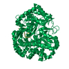
- PDB-9i7w: Extended and wrapped protein P7 dimers of dimers, the P1 layer an... -
+ Open data

- Basic information
| Entry | Database: PDB / ID: 9i7w | ||||||
|---|---|---|---|---|---|---|---|
| Title | Extended and wrapped protein P7 dimers of dimers, the P1 layer and the RNA-dependent RNA polymerase P2 in transcribing particles of bacteriophage phi6 | ||||||
 Components Components |
| ||||||
 Keywords Keywords | VIRAL PROTEIN / inner protein capsid / transcribing phi6 particle / localized reconstruction | ||||||
| Function / homology |  Function and homology information Function and homology informationRNA uridylyltransferase activity / T=2 icosahedral viral capsid / viral inner capsid / virion component / viral nucleocapsid / RNA-directed RNA polymerase / viral RNA genome replication / nucleotide binding / RNA-directed RNA polymerase activity / DNA-templated transcription ...RNA uridylyltransferase activity / T=2 icosahedral viral capsid / viral inner capsid / virion component / viral nucleocapsid / RNA-directed RNA polymerase / viral RNA genome replication / nucleotide binding / RNA-directed RNA polymerase activity / DNA-templated transcription / RNA binding / metal ion binding / identical protein binding Similarity search - Function | ||||||
| Biological species |  Cystovirus phi6 Cystovirus phi6synthetic construct (others) | ||||||
| Method | ELECTRON MICROSCOPY / single particle reconstruction / cryo EM / Resolution: 4.8 Å | ||||||
 Authors Authors | Kumpula, E.-P. / Ilca, S.L. / Huiskonen, J.T. | ||||||
| Funding support |  Finland, 1items Finland, 1items
| ||||||
 Citation Citation | Journal: Mol Cell / Year: 2026 Title: Capsid restructuring activates semi-conservative dsRNA transcription in cystovirus ɸ6. Authors: Serban L Ilca / Xiaoyu Sun / Esa-Pekka Kumpula / Katri Eskelin / David I Stuart / Minna M Poranen / Juha T Huiskonen /  Abstract: Double-stranded (ds)RNA viruses replicate and transcribe their genome within a proteinaceous viral capsid to evade host cell defenses. While Reovirales members use conservative transcription, most ...Double-stranded (ds)RNA viruses replicate and transcribe their genome within a proteinaceous viral capsid to evade host cell defenses. While Reovirales members use conservative transcription, most dsRNA viruses, including cystoviruses, utilize semi-conservative transcription, in which a newly synthesized positive strand replaces the parental positive strand, which is released as mRNA. Here, we visualize semi-conservative transcription activation in cystovirus ɸ6 double-layered particles using cryogenic electron microscopy. We observe nucleotide-triggered disassembly of the domain-swapped outer capsid layer, subsequent expansion of the inner capsid layer, and stepwise assembly of transcription complexes at the opposing poles of the spooled dsRNA genome. These complexes consist of the viral polymerases embedded into a triskelion formed by the minor protein P7, which we show as essential for continuous transcription. The packaging hexamers proximal to the transcription sites channel the viral mRNA exit. Our results define the complex molecular pathway from the quiescent state to activated semi-conservative transcription. | ||||||
| History |
|
- Structure visualization
| Structure viewer | Molecule: MolmilJmol/JSmol |
|---|
- Downloads & links
-Download
| PDBx/mmCIF format | 9i7w.cif.gz | 783.1 KB | Display | PDBx/mmCIF format |
|---|---|---|---|---|
| PDB format | pdb9i7w.ent.gz | 652.2 KB | Display | PDB format |
| PDBx/mmJSON format | 9i7w.json.gz | Tree view | PDBx/mmJSON format | |
| Others |  Other downloads Other downloads |
-Validation report
| Arichive directory | https://data.pdbj.org/pub/pdb/validation_reports/i7/9i7wftp://data.pdbj.org/pub/pdb/validation_reports/i7/9i7w | HTTPS FTP |
|---|
-Related structure data
| Related structure data |  52389MC  9htwC  9hu4C  9hu5C  9hu6C  9i5cC C: citing same article ( M: map data used to model this data |
|---|---|
| Similar structure data |


-Links
 PDBj
PDBj
- Assembly
| Deposited unit | 
|
|---|---|
| 1 |
|
-Components
| #1: DNA chain | Mass: 1445.985 Da / Num. of mol.: 1 / Source method: obtained synthetically / Source: (synth.) synthetic construct (others) | ||||
|---|---|---|---|---|---|
| #2: Protein | Mass: 74903.203 Da / Num. of mol.: 1 / Source method: isolated from a natural source / Source: (natural) Cystovirus phi6 / References: UniProt: P11124, RNA-directed RNA polymerase | ||||
| #3: Protein | Mass: 12017.965 Da / Num. of mol.: 8 / Source method: isolated from a natural source / Source: (natural) Cystovirus phi6 / References: UniProt: Q283U0#4: Protein | Mass: 84783.359 Da / Num. of mol.: 4 / Source method: isolated from a natural source / Source: (natural) Cystovirus phi6 / References: UniProt: P11126Has protein modification | N | |
-Experimental details
-Experiment
| Experiment | Method: ELECTRON MICROSCOPY |
|---|---|
| EM experiment | Aggregation state: PARTICLE / 3D reconstruction method: single particle reconstruction |
- Sample preparation
| Component | Name: Cystovirus phi6 / Type: VIRUS / Entity ID: all / Source: NATURAL | ||||||||||||
|---|---|---|---|---|---|---|---|---|---|---|---|---|---|
| Molecular weight | Experimental value: NO | ||||||||||||
| Source (natural) | Organism: Cystovirus phi6 | ||||||||||||
| Details of virus | Empty: NO / Enveloped: YES / Isolate: SPECIES / Type: VIRION | ||||||||||||
| Virus shell |
| ||||||||||||
| Buffer solution | pH: 8 | ||||||||||||
| Specimen | Embedding applied: NO / Shadowing applied: NO / Staining applied: NO / Vitrification applied: YES | ||||||||||||
| Vitrification | Cryogen name: ETHANE |
- Electron microscopy imaging
| Experimental equipment |  Model: Titan Krios / Image courtesy: FEI Company |
|---|---|
| Microscopy | Model: TFS KRIOS |
| Electron gun | Electron source:  FIELD EMISSION GUN / Accelerating voltage: 300 kV / Illumination mode: FLOOD BEAM FIELD EMISSION GUN / Accelerating voltage: 300 kV / Illumination mode: FLOOD BEAM |
| Electron lens | Mode: BRIGHT FIELD / Nominal defocus max: 41000 nm / Nominal defocus min: 2000 nm |
| Image recording | Electron dose: 42 e/Å2 / Film or detector model: FEI FALCON III (4k x 4k) |
- Processing
| EM software | Name: UCSF ChimeraX / Category: model fitting | ||||||||||||||||||||||||||||
|---|---|---|---|---|---|---|---|---|---|---|---|---|---|---|---|---|---|---|---|---|---|---|---|---|---|---|---|---|---|
| CTF correction | Type: PHASE FLIPPING AND AMPLITUDE CORRECTION | ||||||||||||||||||||||||||||
| Symmetry | Point symmetry: C1 (asymmetric) | ||||||||||||||||||||||||||||
| 3D reconstruction | Resolution: 4.8 Å / Resolution method: FSC 0.143 CUT-OFF / Num. of particles: 18230 / Symmetry type: POINT | ||||||||||||||||||||||||||||
| Atomic model building | Protocol: RIGID BODY FIT | ||||||||||||||||||||||||||||
| Atomic model building |
|
