 Movie
Movie Controller

[English] 日本語
 Yorodumi
- PDB-1hht: RNA dependent RNA polymerase from dsRNA bacteriophage phi6 plus t... -
+ Open data

- Basic information
| Entry | Database: PDB / ID: 1hht | ||||||
|---|---|---|---|---|---|---|---|
| Title | RNA dependent RNA polymerase from dsRNA bacteriophage phi6 plus template | ||||||
 Components Components |
| ||||||
 Keywords Keywords | RNA POLYMERASE / VIRAL POLYMERASE | ||||||
| Function / homology |  Function and homology information Function and homology informationRNA uridylyltransferase activity / virion component / RNA-directed RNA polymerase / viral RNA genome replication / nucleotide binding / RNA-directed RNA polymerase activity / DNA-templated transcription / RNA binding / metal ion binding Similarity search - Function | ||||||
| Biological species |  BACTERIOPHAGE PHI-6 (virus) BACTERIOPHAGE PHI-6 (virus) | ||||||
| Method |  X-RAY DIFFRACTION / SYNCHROTRON / MOLECULAR REPLACEMENT / Resolution: 2.9 Å X-RAY DIFFRACTION / SYNCHROTRON / MOLECULAR REPLACEMENT / Resolution: 2.9 Å | ||||||
 Authors Authors | Grimes, J.M. / Butcher, S.J. / Makeyev, E.V. / Bamford, D.H. / Stuart, D.I. | ||||||
 Citation Citation | Journal: Nature / Year: 2001 Title: A Mechanism for Initiating RNA-Dependent RNA Polymerization Authors: Butcher, S.J. / Grimes, J.M. / Makeyev, E.V. / Bamford, D.H. / Stuart, D.I. #1: Journal: Acta Crystallogr.,Sect.D / Year: 2000 Title: Crystallization and Preliminary X-Ray Crystallographic Studies on the Bacteriophage Phi6 RNA-Dependent RNA Polymerase Authors: Butcher, S.J. / Makeyev, E.V. / Grimes, J.M. / Stuart, D.I. / Bamford, D.H. | ||||||
| History |
|
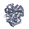
- Structure visualization
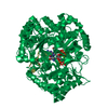
| Structure viewer | Molecule: MolmilJmol/JSmol |
|---|
- Downloads & links
-Download
| PDBx/mmCIF format | 1hht.cif.gz | 396.7 KB | Display | PDBx/mmCIF format |
|---|---|---|---|---|
| PDB format | pdb1hht.ent.gz | 324.2 KB | Display | PDB format |
| PDBx/mmJSON format | 1hht.json.gz | Tree view | PDBx/mmJSON format | |
| Others |  Other downloads Other downloads |
-Validation report
| Arichive directory | https://data.pdbj.org/pub/pdb/validation_reports/hh/1hhtftp://data.pdbj.org/pub/pdb/validation_reports/hh/1hht | HTTPS FTP |
|---|
-Related structure data

-Links
 PDBj
PDBj
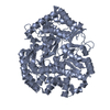
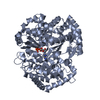
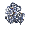
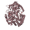
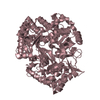
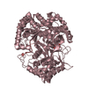
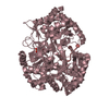
- Assembly
| Deposited unit | 
| ||||||||||||
|---|---|---|---|---|---|---|---|---|---|---|---|---|---|
| 1 | 
| ||||||||||||
| 2 | 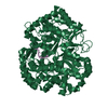
| ||||||||||||
| 3 | 
| ||||||||||||
| Unit cell |
| ||||||||||||
| Noncrystallographic symmetry (NCS) | NCS oper:
| ||||||||||||
| Details | THE PROTEIN IS ACTIVE AS A MONOMER |
-Components
| #1: DNA chain | Mass: 1445.985 Da / Num. of mol.: 3 / Source method: obtained synthetically / Details: 5 NUCLEOTIDE DNA VERSION OF OPTIMUM RNA TEMPLATE #2: Protein | Mass: 74903.203 Da / Num. of mol.: 3 Source method: isolated from a genetically manipulated source Source: (gene. exp.) BACTERIOPHAGE PHI-6 (virus) / Production host:  #3: Chemical |   Mass: 54.938 Da / Num. of mol.: 3 / Source method: obtained synthetically / Formula: Mn Mass: 54.938 Da / Num. of mol.: 3 / Source method: obtained synthetically / Formula: MnCompound details | P2 IS ONE OF THE 4 STRUCTURAL PROTEINS OF THE POLYHEDRAL PROCAPSID. IT IS RESPONSIBLE FOR GENOMIC ...P2 IS ONE OF THE 4 STRUCTURAL | Has protein modification | N | |
|---|
-Experimental details
-Experiment
| Experiment | Method: X-RAY DIFFRACTION / Number of used crystals: 1 |
|---|
- Sample preparation
| Crystal | Density Matthews: 2.92 Å3/Da / Density % sol: 59 % | ||||||||||||||||||||
|---|---|---|---|---|---|---|---|---|---|---|---|---|---|---|---|---|---|---|---|---|---|
| Crystal grow | pH: 6.7 Details: 1:1 PROTEIN(5MG/ML)+TEMPLATE WELL SOLUTION: 10% PEG 8000, 0.1M MES, 2 MM MNCL2, pH 6.70 | ||||||||||||||||||||
| Crystal grow | *PLUS Temperature: 293 K / Method: vapor diffusion, sitting dropDetails: Butcher, S.J., (2000) Acta Crystallogr.,Sect.D, 56, 1473. | ||||||||||||||||||||
| Components of the solutions | *PLUS
|
-Data collection
| Diffraction | Mean temperature: 100 K |
|---|---|
| Diffraction source | Source: SYNCHROTRON / Site: ESRF  / Beamline: BM14 / Wavelength: 0.9724 / Beamline: BM14 / Wavelength: 0.9724 |
| Detector | Type: MARRESEARCH / Detector: CCD / Date: Jun 15, 2000 / Details: TOROIDAL MIRROR |
| Radiation | Monochromator: SI / Protocol: SINGLE WAVELENGTH / Monochromatic (M) / Laue (L): M / Scattering type: x-ray |
| Radiation wavelength | Wavelength: 0.9724 Å / Relative weight: 1 |
| Reflection | Resolution: 2.9→20 Å / Num. obs: 56000 / % possible obs: 95 % / Redundancy: 6.9 % / Rmerge(I) obs: 0.17 / Net I/σ(I): 7.1 |
| Reflection shell | Resolution: 2.9→3 Å / Rmerge(I) obs: 0.96 / Mean I/σ(I) obs: 1.1 / % possible all: 86 |
| Reflection shell | *PLUS % possible obs: 86 % |
- Processing
| Software |
| ||||||||||||||||||||||||||||||||||||||||||||||||||||||||||||||||||||||||||||||||
|---|---|---|---|---|---|---|---|---|---|---|---|---|---|---|---|---|---|---|---|---|---|---|---|---|---|---|---|---|---|---|---|---|---|---|---|---|---|---|---|---|---|---|---|---|---|---|---|---|---|---|---|---|---|---|---|---|---|---|---|---|---|---|---|---|---|---|---|---|---|---|---|---|---|---|---|---|---|---|---|---|---|
| Refinement | Method to determine structure: MOLECULAR REPLACEMENT Starting model: PDB ENTRY TBA Resolution: 2.9→20 Å / Data cutoff high absF: 0 / Isotropic thermal model: RESTRAINED / Cross valid method: THROUGHOUT / σ(F): 0
| ||||||||||||||||||||||||||||||||||||||||||||||||||||||||||||||||||||||||||||||||
| Solvent computation | Solvent model: FLAT MODEL / Bsol: 22 Å2 / ksol: 0.316 e/Å3 | ||||||||||||||||||||||||||||||||||||||||||||||||||||||||||||||||||||||||||||||||
| Displacement parameters | Biso mean: 51 Å2
| ||||||||||||||||||||||||||||||||||||||||||||||||||||||||||||||||||||||||||||||||
| Refine analyze | Luzzati d res low obs: 20 Å | ||||||||||||||||||||||||||||||||||||||||||||||||||||||||||||||||||||||||||||||||
| Refinement step | Cycle: LAST / Resolution: 2.9→20 Å
| ||||||||||||||||||||||||||||||||||||||||||||||||||||||||||||||||||||||||||||||||
| Refine LS restraints |
| ||||||||||||||||||||||||||||||||||||||||||||||||||||||||||||||||||||||||||||||||
| Xplor file |
|
