 Movie
Movie Controller

[English] 日本語
 Yorodumi
- EMDB-52385: Localized reconstruction consensus map of minor protein P7 from t... -
+ Open data

- Basic information
| Entry |  | |||||||||
|---|---|---|---|---|---|---|---|---|---|---|
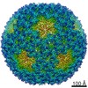
| Title | Localized reconstruction consensus map of minor protein P7 from transcribing particles of bacteriophage phi6 | |||||||||
 Map data Map data | ||||||||||
 Sample Sample |
| |||||||||
 Keywords Keywords | cystovirus / phi6 / semi-conservative transcription / cryo-EM / cryogenic electron microscopy / localized reconstruction / symmetry relaxation / virus / dsRNA virus / bacteriophage | |||||||||
| Function / homology | viral nucleocapsid / Nucleocapsid protein Function and homology information Function and homology information | |||||||||
| Biological species |  Cystovirus phi6 Cystovirus phi6 | |||||||||
| Method | single particle reconstruction / cryo EM / Resolution: 4.1 Å | |||||||||
 Authors Authors | Ilca SL / Huiskonen JT | |||||||||
| Funding support |  Finland, 1 items Finland, 1 items
| |||||||||
 Citation Citation | Journal: Mol Cell / Year: 2026 Title: Capsid restructuring activates semi-conservative dsRNA transcription in cystovirus ɸ6. Authors: Serban L Ilca / Xiaoyu Sun / Esa-Pekka Kumpula / Katri Eskelin / David I Stuart / Minna M Poranen / Juha T Huiskonen /  Abstract: Double-stranded (ds)RNA viruses replicate and transcribe their genome within a proteinaceous viral capsid to evade host cell defenses. While Reovirales members use conservative transcription, most ...Double-stranded (ds)RNA viruses replicate and transcribe their genome within a proteinaceous viral capsid to evade host cell defenses. While Reovirales members use conservative transcription, most dsRNA viruses, including cystoviruses, utilize semi-conservative transcription, in which a newly synthesized positive strand replaces the parental positive strand, which is released as mRNA. Here, we visualize semi-conservative transcription activation in cystovirus ɸ6 double-layered particles using cryogenic electron microscopy. We observe nucleotide-triggered disassembly of the domain-swapped outer capsid layer, subsequent expansion of the inner capsid layer, and stepwise assembly of transcription complexes at the opposing poles of the spooled dsRNA genome. These complexes consist of the viral polymerases embedded into a triskelion formed by the minor protein P7, which we show as essential for continuous transcription. The packaging hexamers proximal to the transcription sites channel the viral mRNA exit. Our results define the complex molecular pathway from the quiescent state to activated semi-conservative transcription. | |||||||||
| History |
|
- Structure visualization
| Supplemental images |
|---|

- Downloads & links
-EMDB archive
| Map data | emd_52385.map.gz | 44.4 MB | EMDB map data format | |
|---|---|---|---|---|
| Header (meta data) | emd-52385-v30.xmlemd-52385.xml | 19.9 KB 19.9 KB | Display Display | EMDB header |
| FSC (resolution estimation) | emd_52385_fsc.xml | 8.3 KB | Display | FSC data file |
| Images |  emd_52385.png emd_52385.png | 168.7 KB | ||
| Masks | emd_52385_msk_1.map | 47.6 MB | Mask map | |
| Filedesc metadata | emd-52385.cif.gz | 5.9 KB | ||
| Others | emd_52385_half_map_1.map.gzemd_52385_half_map_2.map.gz | 36.3 MB 36.3 MB | ||
| Archive directory |  http://ftp.pdbj.org/pub/emdb/structures/EMD-52385ftp://ftp.pdbj.org/pub/emdb/structures/EMD-52385 http://ftp.pdbj.org/pub/emdb/structures/EMD-52385ftp://ftp.pdbj.org/pub/emdb/structures/EMD-52385 | HTTPS FTP |
-Related structure data
| Related structure data |  9htwMC  9hu4C  9hu5C  9hu6C  9i5cC  9i7wC M: atomic model generated by this map C: citing same article ( |
|---|---|
| Similar structure data |


-Links
| EMDB pages | EMDB (EBI/PDBe) / EMDataResource |
|---|
-Map
| File | Download / File: emd_52385.map.gz / Format: CCP4 / Size: 47.6 MB / Type: IMAGE STORED AS FLOATING POINT NUMBER (4 BYTES) | ||||||||||||||||||||
|---|---|---|---|---|---|---|---|---|---|---|---|---|---|---|---|---|---|---|---|---|---|
| Voxel size | X=Y=Z: 1.4 Å | ||||||||||||||||||||
| Density |
| ||||||||||||||||||||
| Symmetry | Space group: 1 | ||||||||||||||||||||
| Details | EMDB XML:
|
-Supplemental data
-Mask #1
| File | emd_52385_msk_1.map | ||||||||||||
|---|---|---|---|---|---|---|---|---|---|---|---|---|---|
| Projections & Slices |
| ||||||||||||
| Density Histograms |
 Z
Z Y
Y X
X


-Half map: #2
| File | emd_52385_half_map_1.map | ||||||||||||
|---|---|---|---|---|---|---|---|---|---|---|---|---|---|
| Projections & Slices |
| ||||||||||||
| Density Histograms |


-Half map: #1
| File | emd_52385_half_map_2.map | ||||||||||||
|---|---|---|---|---|---|---|---|---|---|---|---|---|---|
| Projections & Slices |
| ||||||||||||
| Density Histograms |


- Sample components
-Entire : Cystovirus phi6
| Entire | Name: Cystovirus phi6 |
|---|---|
| Components |
|
-Supramolecule #1: Cystovirus phi6
| Supramolecule | Name: Cystovirus phi6 / type: virus / ID: 1 / Parent: 0 / Macromolecule list: all / NCBI-ID: 10879 / Sci species name: Cystovirus phi6 / Virus type: VIRION / Virus isolate: SPECIES / Virus enveloped: Yes / Virus empty: No |
|---|---|
| Virus shell | Shell ID: 1 / Name: Outer protein shell / T number (triangulation number): 13 |
| Virus shell | Shell ID: 2 / Name: Inner protein shell / T number (triangulation number): 1 |
-Macromolecule #1: Nucleocapsid protein
| Macromolecule | Name: Nucleocapsid protein / type: protein_or_peptide / ID: 1 / Number of copies: 2 / Enantiomer: LEVO |
|---|---|
| Source (natural) | Organism: Cystovirus phi6 |
| Molecular weight | Theoretical: 12.017965 KDa |
| Sequence | String: TLYLVPPLDS ADKELPALAS KAGVTLLEIE FLHELWPHLS GGQIVIAALN ANNLAILNRH MSTLLVELPV AVMAVPGASY RSDWNMIAH ALPSEDWITL SNKMLKSGLL AN UniProtKB: Nucleocapsid protein |
-Experimental details
-Structure determination
| Method | cryo EM |
|---|---|
 Processing Processing | single particle reconstruction |
| Aggregation state | particle |
-Sample preparation
| Buffer | pH: 8 |
|---|---|
| Vitrification | Cryogen name: ETHANE |
- Electron microscopy
| Microscope | TFS KRIOS |
|---|---|
| Image recording | Film or detector model: FEI FALCON III (4k x 4k) / Average electron dose: 42.0 e/Å2 |
| Electron beam | Acceleration voltage: 300 kV / Electron source:  FIELD EMISSION GUN FIELD EMISSION GUN |
| Electron optics | Illumination mode: FLOOD BEAM / Imaging mode: BRIGHT FIELD / Nominal defocus max: 41.0 µm / Nominal defocus min: 2.0 µm |
| Experimental equipment |  Model: Titan Krios / Image courtesy: FEI Company |
+Image processing
-Atomic model buiding 1
| Initial model | Chain - Source name: AlphaFold / Chain - Initial model type: in silico model |
|---|---|
| Output model | PDB-9htw: |
