- PDB-9i24: Coagulation factor Xa complex with a2-loop peptide -
+
Open data
ID or keywords:
Loading...
-
Basic information
Entry
Database: PDB / ID: 9i24
Title
Coagulation factor Xa complex with a2-loop peptide
Components
Activated factor Xa heavy chain
Coagulation factor V heavy chain
Factor X light chain
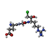
GLU-GLY-ARG-chloromethyl ketone inhibitor (EGRCK)
Keywords
BLOOD CLOTTING / protease / enzyme / complex
Function / homology
Function and homology information
coagulation factor Xa / Defective factor IX causes thrombophilia / Defective cofactor function of FVIIIa variant / Defective F9 variant does not activate FX / : / positive regulation of TOR signaling / Transport of gamma-carboxylated protein precursors from the endoplasmic reticulum to the Golgi apparatus / : / Gamma-carboxylation of protein precursors / Removal of aminoterminal propeptides from gamma-carboxylated proteins ...coagulation factor Xa / Defective factor IX causes thrombophilia / Defective cofactor function of FVIIIa variant / Defective F9 variant does not activate FX / : / positive regulation of TOR signaling / Transport of gamma-carboxylated protein precursors from the endoplasmic reticulum to the Golgi apparatus / : / Gamma-carboxylation of protein precursors / Removal of aminoterminal propeptides from gamma-carboxylated proteins / : / phospholipid binding / Golgi lumen / blood coagulation / positive regulation of cell migration / endoplasmic reticulum lumen / serine-type endopeptidase activity / external side of plasma membrane / calcium ion binding / proteolysis / : / extracellular region / plasma membrane Similarity search - Function
Journal: Blood / Year: 2026 Title: A 3.3-Å cryo-EM structure of an engineered high-affinity human prothrombinase complex. Authors: Fatma Işık Üstok / Alexandre Faille / James A Huntington / Abstract: Thrombin is generated from prothrombin through cleavage at 2 sites by the enzyme prothrombinase, composed of factor Xa (fXa) and fVa. The affinity of fXa for fVa is low, with assembly and function ...Thrombin is generated from prothrombin through cleavage at 2 sites by the enzyme prothrombinase, composed of factor Xa (fXa) and fVa. The affinity of fXa for fVa is low, with assembly and function dependent on phospholipid (PL) membranes. Some snakes have evolved venom versions of fXa that bind to fVa with high affinity and efficiently activate prothrombin in the absence of PL. We created a similar high-affinity, PL-independent human prothrombinase with 17 mutations to human fXa (M17). The increase in affinity enabled cryogenic electron microscopy (cryo-EM) structure determination of M17-prothrombinase to a resolution of 3.3 Å. All protein domains were well resolved in the map, except for the γ-carboxyglutamic acid domain of fXa. The main contacts involve the serine protease and epidermal growth factor-like domain 2 (EGF2) domains of fXa and the A2 and A3 domains of fVa, resulting in the burying of a total surface area of 4900 Å2. The map is of sufficient quality to resolve side-chain interactions, including several key M17 mutations. To aid in the placement of the loop C-terminal to the A2 domain (a2-loop), we solved a high-resolution crystal structure of fXa in complex with a synthetic a2 peptide. The acidic a2-loop interacts with the basic heparin-binding site of fXa, involving a conserved antiparallel β-strand interaction. The M17-prothrombinase structure is compatible with data from biochemical and mutagenesis research and provides important new insights into the assembly and function of the prothrombinase complex.
#259 - Jul 2021 Designed Proteins and Citizen Science similarity (1)
-
Assembly
Deposited unit
H: Activated factor Xa heavy chain L: Factor X light chain C: Coagulation factor V heavy chain I: GLU-GLY-ARG-chloromethyl ketone inhibitor (EGRCK) hetero molecules
Mass: 28538.541 Da / Num. of mol.: 1 Source method: isolated from a genetically manipulated source Source: (gene. exp.) Homo sapiens (human) / Gene: F10 / Production host: Escherichia coli (E. coli) / References: UniProt: P00742
#2: Protein
FactorXlightchain
Mass: 6034.818 Da / Num. of mol.: 1 Source method: isolated from a genetically manipulated source Source: (gene. exp.) Homo sapiens (human) / Gene: F10 / Production host: Escherichia coli (E. coli) / References: UniProt: P00742
-
Protein/peptide , 2 types, 2 molecules CI
#3: Protein/peptide
CoagulationfactorVheavychain
Mass: 1866.800 Da / Num. of mol.: 1 / Source method: obtained synthetically Details: The Y is phosphorylated (PTR); the N-terminus is acetylated; the C-terminus is amidated. Source: (synth.) synthetic construct (others)
#4: Protein/peptide
GLU-GLY-ARG-chloromethylketoneinhibitor (EGRCK)
Mass: 361.375 Da / Num. of mol.: 1 / Source method: obtained synthetically Details: The R is modified and the identifier OGJ is used. It is covalently linked to the catalytic Ser195 and His57. Source: (synth.) synthetic construct (others)
In the structure databanks used in Yorodumi, some data are registered as the other names, "COVID-19 virus" and "2019-nCoV". Here are the details of the virus and the list of structure data.
Jan 31, 2019. EMDB accession codes are about to change! (news from PDBe EMDB page)
EMDB accession codes are about to change! (news from PDBe EMDB page)
The allocation of 4 digits for EMDB accession codes will soon come to an end. Whilst these codes will remain in use, new EMDB accession codes will include an additional digit and will expand incrementally as the available range of codes is exhausted. The current 4-digit format prefixed with “EMD-” (i.e. EMD-XXXX) will advance to a 5-digit format (i.e. EMD-XXXXX), and so on. It is currently estimated that the 4-digit codes will be depleted around Spring 2019, at which point the 5-digit format will come into force.
The EM Navigator/Yorodumi systems omit the EMD- prefix.
Related info.:Q: What is EMD? / ID/Accession-code notation in Yorodumi/EM Navigator
Yorodumi is a browser for structure data from EMDB, PDB, SASBDB, etc.
This page is also the successor to EM Navigator detail page, and also detail information page/front-end page for Omokage search.
The word "yorodu" (or yorozu) is an old Japanese word meaning "ten thousand". "mi" (miru) is to see.
Related info.:EMDB / PDB / SASBDB / Comparison of 3 databanks / Yorodumi Search / Aug 31, 2016. New EM Navigator & Yorodumi / Yorodumi Papers / Jmol/JSmol / Function and homology information / Changes in new EM Navigator and Yorodumi
 Movie
Movie Controller


 Open data
 Basic information
 Components
 Keywords
 Function and homology information
 Homo sapiens (human)
 X-RAY DIFFRACTION /
  Authors
 United Kingdom, 1items
  Citation
 Structure visualization
 Downloads & links
 Other downloads

 PDBj
PDBj


 Assembly


 Mass: 40.078 Da / Num. of mol.: 1 / Source method: obtained synthetically / Formula: Ca
 Mass: 22.990 Da / Num. of mol.: 1 / Source method: obtained synthetically / Formula: Na
 Mass: 35.453 Da / Num. of mol.: 1 / Source method: obtained synthetically / Formula: Cl
 Mass: 69.085 Da / Num. of mol.: 1 / Source method: obtained synthetically / Formula: C3H5N2
 Type: peptide-like / Mass: 395.862 Da / Num. of mol.: 1 / Source method: obtained synthetically / Formula: C14H28ClN6O5
 Sample preparation
 Processing