 Movie
Movie Controller

[English] 日本語
 Yorodumi
- PDB-9hfs: Cryo-EM structure of human CDADC1 inactive mutant (E400A): hexame... -
+ Open data

- Basic information
| Entry | Database: PDB / ID: 9hfs | |||||||||||||||
|---|---|---|---|---|---|---|---|---|---|---|---|---|---|---|---|---|
| Title | Cryo-EM structure of human CDADC1 inactive mutant (E400A): hexamer with dCTP bound in the active site | |||||||||||||||
 Components Components | Cytidine and dCMP deaminase domain-containing protein 1 | |||||||||||||||
 Keywords Keywords | HYDROLASE / Human dCTP deaminase / trimer / Zinc-dependent / Nucleotide metabolism | |||||||||||||||
| Function / homology |  Function and homology information Function and homology informationdCMP deaminase activity / dCTP deaminase / DNA cytosine deamination / cytidine deaminase activity / importin-alpha family protein binding / protein homodimerization activity / zinc ion binding / nucleus / cytoplasm Similarity search - Function | |||||||||||||||
| Biological species |  Homo sapiens (human) Homo sapiens (human) | |||||||||||||||
| Method | ELECTRON MICROSCOPY / single particle reconstruction / cryo EM / Resolution: 2.8 Å | |||||||||||||||
 Authors Authors | Slyvka, A. / Rathore, I. / Yang, R. / Kanai, T. / Lountos, G. / Wang, Z. / Skowronek, K. / Czarnocki-Cieciura, M. / Wlodawer, A. / Bochtler, M. | |||||||||||||||
| Funding support |  Poland, Poland,  United States, 4items United States, 4items
| |||||||||||||||
 Citation Citation | Journal: Proc Natl Acad Sci U S A / Year: 2025 Title: Activity and structure of human (d)CTP deaminase CDADC1. Authors: Anton Slyvka / Ishan Rathore / Renbin Yang / Olga Gewartowska / Tapan Kanai / George T Lountos / Krzysztof Skowronek / Mariusz Czarnocki-Cieciura / Alexander Wlodawer / Matthias Bochtler / Abstract: Vertebrates have evolved an understudied protein termed CDADC1 (NYD-SP15) that contains an inactive N-terminal and active C-terminal DCTD-like domain. Here, we show that human CDADC1 is a (d)CTP- ...Vertebrates have evolved an understudied protein termed CDADC1 (NYD-SP15) that contains an inactive N-terminal and active C-terminal DCTD-like domain. Here, we show that human CDADC1 is a (d)CTP-specific deaminase, with a roughly 2-fold in vitro preference for dCTP over CTP. We determined high-resolution cryo-EM structures of CDADC1 in the absence of substrate and in complex with dCTP and 5-methyl-dCTP. The structures show that CDADC1 forms trimers and dimers of trimers in solution. The (d)CTP substrate is selected by a narrow pocket for the cytosine base and multiple lysine and arginine contacts to the triphosphate. Substrate binding promotes the association of trimers into hexamers and the transition of the hexamers from a loose to a tighter arrangement. Genetic experiments in mice show that loss of Cdadc1 is surprisingly well tolerated, even in the absence of the dCMP deaminase Dctd that is considered as the main source of dUMP, the precursor of dTTP. | |||||||||||||||
| History |
|
- Structure visualization
| Structure viewer | Molecule: MolmilJmol/JSmol |
|---|
- Downloads & links
-Download
| PDBx/mmCIF format | 9hfs.cif.gz | 576 KB | Display | PDBx/mmCIF format |
|---|---|---|---|---|
| PDB format | pdb9hfs.ent.gz | 373 KB | Display | PDB format |
| PDBx/mmJSON format | 9hfs.json.gz | Tree view | PDBx/mmJSON format | |
| Others |  Other downloads Other downloads |
-Validation report
| Arichive directory | https://data.pdbj.org/pub/pdb/validation_reports/hf/9hfsftp://data.pdbj.org/pub/pdb/validation_reports/hf/9hfs | HTTPS FTP |
|---|
-Related structure data
| Related structure data |  52124MC 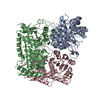 9hfqC  9hfrC  9hftC C: citing same article ( M: map data used to model this data |
|---|---|
| Similar structure data |


-Links
 PDBj
PDBj- Assembly
| Deposited unit | 
|
|---|---|
| 1 |
|
-Components
| #1: Protein | Mass: 60652.320 Da / Num. of mol.: 6 / Mutation: E400A Source method: isolated from a genetically manipulated source Source: (gene. exp.) Homo sapiens (human) / Gene: CDADC1 / Production host:  #2: Chemical | ChemComp-ZN /   Mass: 65.409 Da / Num. of mol.: 12 / Source method: obtained synthetically / Formula: Zn / Feature type: SUBJECT OF INVESTIGATION Mass: 65.409 Da / Num. of mol.: 12 / Source method: obtained synthetically / Formula: Zn / Feature type: SUBJECT OF INVESTIGATION#3: Chemical | ChemComp-DCP /   Mass: 467.157 Da / Num. of mol.: 6 / Source method: obtained synthetically / Formula: C9H16N3O13P3 / Feature type: SUBJECT OF INVESTIGATION Mass: 467.157 Da / Num. of mol.: 6 / Source method: obtained synthetically / Formula: C9H16N3O13P3 / Feature type: SUBJECT OF INVESTIGATION#4: Water | ChemComp-HOH / |  Mass: 18.015 Da / Num. of mol.: 6 / Source method: isolated from a natural source / Formula: H2O Mass: 18.015 Da / Num. of mol.: 6 / Source method: isolated from a natural source / Formula: H2OHas ligand of interest | Y | Has protein modification | N | |
|---|
-Experimental details
-Experiment
| Experiment | Method: ELECTRON MICROSCOPY |
|---|---|
| EM experiment | Aggregation state: PARTICLE / 3D reconstruction method: single particle reconstruction |
- Sample preparation
| Component | Name: Human CDADC1 hexamer with bound dCTP / Type: COMPLEX / Entity ID: #1 / Source: RECOMBINANT |
|---|---|
| Molecular weight | Value: 0.363 MDa / Experimental value: YES |
| Source (natural) | Organism: Homo sapiens (human) |
| Source (recombinant) | Organism: |
| Buffer solution | pH: 7.4 |
| Specimen | Conc.: 0.7 mg/ml / Embedding applied: NO / Shadowing applied: NO / Staining applied: NO / Vitrification applied: YES |
| Specimen support | Grid material: COPPER / Grid mesh size: 200 divisions/in. / Grid type: Quantifoil R1.2/1.3 |
| Vitrification | Instrument: FEI VITROBOT MARK IV / Cryogen name: ETHANE / Humidity: 100 % / Chamber temperature: 277.15 K |
- Electron microscopy imaging
| Experimental equipment |  Model: Talos Arctica / Image courtesy: FEI Company |
|---|---|
| Microscopy | Model: FEI TALOS ARCTICA |
| Electron gun | Electron source:  FIELD EMISSION GUN / Accelerating voltage: 200 kV / Illumination mode: FLOOD BEAM FIELD EMISSION GUN / Accelerating voltage: 200 kV / Illumination mode: FLOOD BEAM |
| Electron lens | Mode: BRIGHT FIELD / Nominal defocus max: 2500 nm / Nominal defocus min: 800 nm |
| Specimen holder | Cryogen: NITROGEN |
| Image recording | Electron dose: 42.6 e/Å2 / Film or detector model: GATAN K3 BIOQUANTUM (6k x 4k) |
- Processing
| EM software |
| |||||||||||||||||||||||||||||||||
|---|---|---|---|---|---|---|---|---|---|---|---|---|---|---|---|---|---|---|---|---|---|---|---|---|---|---|---|---|---|---|---|---|---|---|
| CTF correction | Type: PHASE FLIPPING AND AMPLITUDE CORRECTION | |||||||||||||||||||||||||||||||||
| Symmetry | Point symmetry: D3 (2x3 fold dihedral) | |||||||||||||||||||||||||||||||||
| 3D reconstruction | Resolution: 2.8 Å / Resolution method: FSC 0.143 CUT-OFF / Num. of particles: 310460 / Symmetry type: POINT | |||||||||||||||||||||||||||||||||
| Atomic model building | Protocol: AB INITIO MODEL / Space: REAL | |||||||||||||||||||||||||||||||||
| Atomic model building | Source name: AlphaFold / Type: in silico model | |||||||||||||||||||||||||||||||||
| Refinement | Cross valid method: NONE Stereochemistry target values: GeoStd + Monomer Library + CDL v1.2 | |||||||||||||||||||||||||||||||||
| Displacement parameters | Biso mean: 19.51 Å2 | |||||||||||||||||||||||||||||||||
| Refine LS restraints |
|
