 Movie
Movie Controller

[English] 日本語
 Yorodumi
- PDB-9h19: Cryo-EM structure of RC-dLH complex model I from Gem. groenlandic... -
+ Open data

- Basic information
| Entry | Database: PDB / ID: 9h19 | |||||||||||||||||||||||||||
|---|---|---|---|---|---|---|---|---|---|---|---|---|---|---|---|---|---|---|---|---|---|---|---|---|---|---|---|---|
| Title | Cryo-EM structure of RC-dLH complex model I from Gem. groenlandica strain TET16 | |||||||||||||||||||||||||||
 Components Components |
| |||||||||||||||||||||||||||
 Keywords Keywords | PHOTOSYNTHESIS / reaction centre light harvesting complex / RC-dLH / photosynthetic bacteria / gemmatimonas groenlandica | |||||||||||||||||||||||||||
| Function / homology |  Function and homology information Function and homology informationbacteriochlorophyll binding / organelle inner membrane / plasma membrane light-harvesting complex / photosynthetic electron transport in photosystem II / photosynthesis, light reaction / endomembrane system / electron transfer activity / iron ion binding / heme binding / metal ion binding / plasma membrane Similarity search - Function | |||||||||||||||||||||||||||
| Biological species |  Gemmatimonas groenlandica (bacteria) Gemmatimonas groenlandica (bacteria) | |||||||||||||||||||||||||||
| Method | ELECTRON MICROSCOPY / single particle reconstruction / cryo EM / Resolution: 2.3 Å | |||||||||||||||||||||||||||
 Authors Authors | Gardiner, A. / Qian, P. / Koblizek, M. / Jing, Y. / Joosten, M. / Jakobi, A. / Bina, D. / Mujakic, I. / Gardian, Z. / Kaftan, D. / Castro-Hartmann, P. | |||||||||||||||||||||||||||
| Funding support |  Czech Republic, 1items Czech Republic, 1items
| |||||||||||||||||||||||||||
 Citation Citation | Journal: mSystems / Year: 2026 Title: Two solutions for efficient light-harvesting in phototrophic . Authors: Alastair T Gardiner / Yibo Jin / David Bína / Maarten Joosten / David Kaftan / Izabela Mujakić / Zdenko Gardian / Pablo Castro-Hartmann / Pu Qian / Michal Koblížek /   Abstract: Phototrophic Gemmatimonadota represent a unique group of phototrophic bacteria that acquired a complete set of photosynthetic genes via horizontal gene transfer and later evolved independently. () ...Phototrophic Gemmatimonadota represent a unique group of phototrophic bacteria that acquired a complete set of photosynthetic genes via horizontal gene transfer and later evolved independently. () contains photosynthetic complexes with two concentric light-harvesting antenna rings that absorb at 816 and 868 nm, allowing it to better exploit the light conditions found deeper in the water column. The closely related species , with highly similar photosynthetic genes, harvests infrared light using a single 860 nm absorption band. The cryo-electron microscopy structure of the photosynthetic complex reveals that the outer antenna lacks monomeric bacteriochlorophylls, resulting in a smaller optical antenna cross-section. The spectrum is red-shifted relative to due to the formation of a H-bond enabled by a different rotamer conformation of αTrp in the outer ring. This H-bond forms with a neighboring bacteriochlorophyll and increases the intra-dimer exciton coupling, affecting the exciton localization probability within the rings and increasing exciton cooperativity between the complexes. The functional consequences of the spectral shift, caused solely by a subtle conformational change of a single residue, represent a novel mechanism in which phototrophic organisms adjust their antennae for particular light conditions and enable to grow higher in the water column where more photons are available.IMPORTANCEThe photoheterotrophic species of the phylum Gemmatimonadota employ unique photosynthetic complexes with two concentric antenna rings around a central reaction center. In contrast to other phototrophic species, these organisms have not evolved any regulatory systems to control the expression of their photosynthetic apparatus under different light conditions. Despite the overall similarity, the complexes present in and have different absorption properties in the near-infrared region of the spectrum that make them more suitable for low or medium light, respectively. The main difference in absorption depends on the conformation of a single tryptophan residue that can form an H-bond with a neighboring bacteriochlorophyll. The presence or absence of this H-bond affects how the protein scaffold interacts with the bacteriochlorophylls, which in turn determines how light energy is transferred within and between the photosynthetic complexes. | |||||||||||||||||||||||||||
| History |
|
- Structure visualization
| Structure viewer | Molecule: MolmilJmol/JSmol |
|---|
- Downloads & links
-Download
| PDBx/mmCIF format | 9h19.cif.gz | 1.5 MB | Display | PDBx/mmCIF format |
|---|---|---|---|---|
| PDB format | pdb9h19.ent.gz | Display | PDB format | |
| PDBx/mmJSON format | 9h19.json.gz | Tree view | PDBx/mmJSON format | |
| Others |  Other downloads Other downloads |
-Validation report
| Arichive directory | https://data.pdbj.org/pub/pdb/validation_reports/h1/9h19ftp://data.pdbj.org/pub/pdb/validation_reports/h1/9h19 | HTTPS FTP |
|---|
-Related structure data
| Related structure data |  51760MC  9h22C M: map data used to model this data C: citing same article ( |
|---|---|
| Similar structure data |

-Links
 PDBj
PDBj
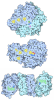
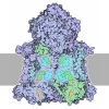
- Assembly
| Deposited unit | 
|
|---|---|
| 1 |
|
-Components
-Light-harvesting ... , 3 types, 80 molecules AaAbAcAdAeAfAgAhAiAjAkAlAmAnAoApBaBbBcBdBeBfBgBhBiBjBkBlBmBn...
| #1: Protein | Mass: 7697.123 Da / Num. of mol.: 16 / Source method: isolated from a natural source / Details: helix / Source: (natural) Gemmatimonas groenlandica (bacteria) / References: UniProt: A0A6M4INU2#2: Protein/peptide | Mass: 5183.985 Da / Num. of mol.: 40 / Source method: isolated from a natural source / Details: helix / Source: (natural) Gemmatimonas groenlandica (bacteria) / References: UniProt: A0A6M4IQ93#9: Protein | Mass: 6413.583 Da / Num. of mol.: 24 / Source method: isolated from a natural source / Details: helix / Source: (natural) Gemmatimonas groenlandica (bacteria) / References: UniProt: A0A6M4IQ17 |
|---|
-Reaction centre ... , 3 types, 3 molecules SHK
| #3: Protein | Mass: 21641.564 Da / Num. of mol.: 1 / Source method: isolated from a natural source / Details: native protein / Source: (natural) Gemmatimonas groenlandica (bacteria) / References: UniProt: A0A6M4IPJ3 |
|---|---|
| #6: Protein | Mass: 7202.296 Da / Num. of mol.: 1 / Source method: isolated from a natural source / Details: helix / Source: (natural) Gemmatimonas groenlandica (bacteria) |
| #7: Protein | Mass: 19538.121 Da / Num. of mol.: 1 / Source method: isolated from a natural source / Details: native proteins / Source: (natural) Gemmatimonas groenlandica (bacteria) |
-Reaction center protein ... , 2 types, 2 molecules LM
| #4: Protein | Mass: 30542.740 Da / Num. of mol.: 1 / Source method: isolated from a natural source / Details: native protein / Source: (natural) Gemmatimonas groenlandica (bacteria) / References: UniProt: A0A6M4IPC7 |
|---|---|
| #5: Protein | Mass: 43396.742 Da / Num. of mol.: 1 / Source method: isolated from a natural source / Details: native proteins / Source: (natural) Gemmatimonas groenlandica (bacteria) / References: UniProt: A0A6M4ILR2 |
-Protein / Sugars , 2 types, 49 molecules C
| #12: Sugar | ChemComp-LMT / 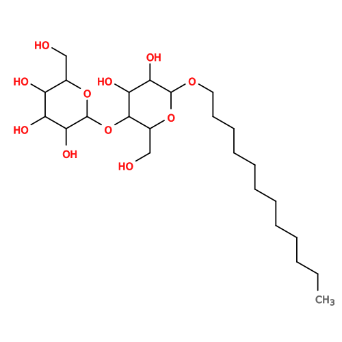 Type: D-saccharide / Mass: 510.615 Da / Num. of mol.: 48 / Source method: obtained synthetically / Formula: C24H46O11 / Comment: detergent*YM Type: D-saccharide / Mass: 510.615 Da / Num. of mol.: 48 / Source method: obtained synthetically / Formula: C24H46O11 / Comment: detergent*YM#8: Protein | | Mass: 40302.758 Da / Num. of mol.: 1 / Source method: isolated from a natural source / Details: native protein / Source: (natural) Gemmatimonas groenlandica (bacteria) / References: UniProt: A0A6M4ITL7 |
|---|
-Non-polymers , 10 types, 192 molecules 


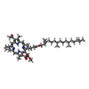

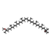
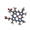

| #10: Chemical | ChemComp-V7N / (  Mass: 610.865 Da / Num. of mol.: 40 / Source method: obtained synthetically / Formula: C41H54O4 Mass: 610.865 Da / Num. of mol.: 40 / Source method: obtained synthetically / Formula: C41H54O4#11: Chemical | ChemComp-BCL / 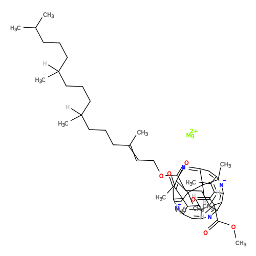 Mass: 911.504 Da / Num. of mol.: 84 / Source method: obtained synthetically / Formula: C55H74MgN4O6 / Feature type: SUBJECT OF INVESTIGATION Mass: 911.504 Da / Num. of mol.: 84 / Source method: obtained synthetically / Formula: C55H74MgN4O6 / Feature type: SUBJECT OF INVESTIGATION#13: Chemical | ChemComp-PEX / 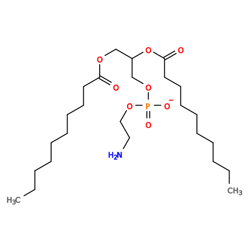 Mass: 522.632 Da / Num. of mol.: 17 / Source method: obtained synthetically / Formula: C25H49NO8P Mass: 522.632 Da / Num. of mol.: 17 / Source method: obtained synthetically / Formula: C25H49NO8P#14: Chemical |  Mass: 717.116 Da / Num. of mol.: 3 / Source method: obtained synthetically / Formula: C51H72O2 Mass: 717.116 Da / Num. of mol.: 3 / Source method: obtained synthetically / Formula: C51H72O2#15: Chemical | 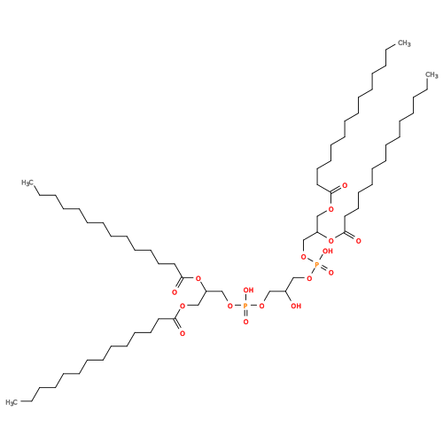 Mass: 1241.633 Da / Num. of mol.: 2 / Source method: obtained synthetically / Formula: C65H126O17P2 Mass: 1241.633 Da / Num. of mol.: 2 / Source method: obtained synthetically / Formula: C65H126O17P2#16: Chemical | 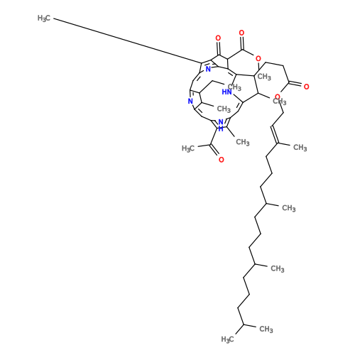 Mass: 889.215 Da / Num. of mol.: 2 / Source method: obtained synthetically / Formula: C55H76N4O6 Mass: 889.215 Da / Num. of mol.: 2 / Source method: obtained synthetically / Formula: C55H76N4O6#17: Chemical | ChemComp-FE / |  Mass: 55.845 Da / Num. of mol.: 1 / Source method: obtained synthetically / Formula: Fe Mass: 55.845 Da / Num. of mol.: 1 / Source method: obtained synthetically / Formula: Fe#18: Chemical | ChemComp-CRT / |  Mass: 596.925 Da / Num. of mol.: 1 / Source method: obtained synthetically / Formula: C42H60O2 Mass: 596.925 Da / Num. of mol.: 1 / Source method: obtained synthetically / Formula: C42H60O2#19: Chemical | ChemComp-HEC /  Mass: 620.519 Da / Num. of mol.: 4 / Source method: obtained synthetically / Formula: C34H36FeN4O4 Mass: 620.519 Da / Num. of mol.: 4 / Source method: obtained synthetically / Formula: C34H36FeN4O4#20: Water | ChemComp-HOH / | Mass: 18.015 Da / Num. of mol.: 38 / Source method: isolated from a natural source / Formula: H2O |
|---|
-Details
| Has ligand of interest | Y |
|---|---|
| Has protein modification | Y |
-Experimental details
-Experiment
| Experiment | Method: ELECTRON MICROSCOPY |
|---|---|
| EM experiment | Aggregation state: PARTICLE / 3D reconstruction method: single particle reconstruction |
- Sample preparation
| Component | Name: RC-dLH complex model I from Gemmatimonas groenlandica / Type: COMPLEX / Details: a single monomer complex / Entity ID: #9, #1-#2, #8, #6-#7, #4-#5, #3 / Source: NATURAL |
|---|---|
| Molecular weight | Value: 0.65 MDa / Experimental value: NO |
| Source (natural) | Organism: Gemmatimonas groenlandica (bacteria) |
| Buffer solution | pH: 8 / Details: 20 mM Tris-HCL, 50 mM NaCl, pH 8.0, 0.02% beta-DDM |
| Buffer component | Conc.: 20 mM / Name: Tris-HCL |
| Specimen | Conc.: 4 mg/ml / Embedding applied: NO / Shadowing applied: NO / Staining applied: NO / Vitrification applied: YES Details: Protein in buffer solution with detergent of beta-DDM |
| Specimen support | Grid material: COPPER / Grid mesh size: 300 divisions/in. / Grid type: Quantifoil R1.2/1.3 |
| Vitrification | Instrument: FEI VITROBOT MARK IV / Cryogen name: ETHANE / Humidity: 100 % / Chamber temperature: 277 K / Details: Blot time 3 seconds, blot force, 3 |
- Electron microscopy imaging
| Experimental equipment |  Model: Titan Krios / Image courtesy: FEI Company |
|---|---|
| Microscopy | Model: TFS KRIOS |
| Electron gun | Electron source:  FIELD EMISSION GUN / Accelerating voltage: 300 kV / Illumination mode: FLOOD BEAM FIELD EMISSION GUN / Accelerating voltage: 300 kV / Illumination mode: FLOOD BEAM |
| Electron lens | Mode: BRIGHT FIELD / Nominal magnification: 165000 X / Nominal defocus max: 2400 nm / Nominal defocus min: 800 nm / Cs: 2.7 mm / C2 aperture diameter: 100 µm / Alignment procedure: COMA FREE |
| Specimen holder | Cryogen: NITROGEN / Specimen holder model: FEI TITAN KRIOS AUTOGRID HOLDER / Temperature (max): 92 K / Temperature (min): 88 K |
| Image recording | Average exposure time: 5.13 sec. / Electron dose: 60 e/Å2 / Film or detector model: FEI FALCON IV (4k x 4k) / Num. of grids imaged: 1 / Num. of real images: 23012 |
| EM imaging optics | Energyfilter name: TFS Selectris X Details: Thermofisher Titan Krios G3 with energy filter selectrisX Energyfilter slit width: 10 eV |
- Processing
| EM software |
| ||||||||||||||||||||||||||||||||||||||||
|---|---|---|---|---|---|---|---|---|---|---|---|---|---|---|---|---|---|---|---|---|---|---|---|---|---|---|---|---|---|---|---|---|---|---|---|---|---|---|---|---|---|
| Image processing | Details: Tiff format was used for image recording. | ||||||||||||||||||||||||||||||||||||||||
| CTF correction | Details: CTF was performed within cryosparc / Type: PHASE FLIPPING AND AMPLITUDE CORRECTION | ||||||||||||||||||||||||||||||||||||||||
| Particle selection | Num. of particles selected: 935540 Details: Initially, particles were selected by the use of cryosparc blob model automatically. Model reference then was used later for final 3D reconstruction. | ||||||||||||||||||||||||||||||||||||||||
| Symmetry | Point symmetry: C1 (asymmetric) | ||||||||||||||||||||||||||||||||||||||||
| 3D reconstruction | Resolution: 2.3 Å / Resolution method: FSC 0.143 CUT-OFF / Num. of particles: 129052 / Algorithm: BACK PROJECTION / Num. of class averages: 1 / Symmetry type: POINT | ||||||||||||||||||||||||||||||||||||||||
| Atomic model building | B value: 60 / Protocol: RIGID BODY FIT / Space: REAL / Target criteria: model to map | ||||||||||||||||||||||||||||||||||||||||
| Atomic model building | Source name: AlphaFold / Type: in silico model |
