 Movie
Movie Controller

[English] 日本語
 Yorodumi
- PDB-9gsi: Cryo-EM structure of mouse PMCA captured in E1-ATP in the presenc... -
+ Open data

- Basic information
| Entry | Database: PDB / ID: 9gsi | |||||||||||||||||||||
|---|---|---|---|---|---|---|---|---|---|---|---|---|---|---|---|---|---|---|---|---|---|---|
| Title | Cryo-EM structure of mouse PMCA captured in E1-ATP in the presence of Calcium | |||||||||||||||||||||
 Components Components | Plasma membrane calcium-transporting ATPase 2 | |||||||||||||||||||||
 Keywords Keywords | MEMBRANE PROTEIN / Calcium pump / Ptype-Atpase | |||||||||||||||||||||
| Function / homology |  Function and homology information Function and homology informationP-type calcium transporter activity involved in regulation of postsynaptic cytosolic calcium ion concentration / cerebellar Purkinje cell layer morphogenesis / otolith mineralization / inner ear receptor cell differentiation / Reduction of cytosolic Ca++ levels / cGMP metabolic process / Ion transport by P-type ATPases / cerebellar granule cell differentiation / cerebellar Purkinje cell differentiation / calcium-dependent ATPase activity ...P-type calcium transporter activity involved in regulation of postsynaptic cytosolic calcium ion concentration / cerebellar Purkinje cell layer morphogenesis / otolith mineralization / inner ear receptor cell differentiation / Reduction of cytosolic Ca++ levels / cGMP metabolic process / Ion transport by P-type ATPases / cerebellar granule cell differentiation / cerebellar Purkinje cell differentiation / calcium-dependent ATPase activity / photoreceptor ribbon synapse / P-type Ca2+ transporter / detection of mechanical stimulus involved in sensory perception of sound / P-type calcium transporter activity / Ion homeostasis / serotonin metabolic process / positive regulation of calcium ion transport / auditory receptor cell stereocilium organization / locomotion / neuromuscular process controlling balance / inner ear morphogenesis / dendritic spine membrane / regulation of cell size / inner ear development / neuronal cell body membrane / parallel fiber to Purkinje cell synapse / cochlea development / glutamate receptor binding / regulation of cytosolic calcium ion concentration / lactation / presynaptic active zone membrane / cerebellum development / PDZ domain binding / sensory perception of sound / locomotory behavior / regulation of synaptic plasticity / synapse organization / postsynaptic density membrane / cell morphogenesis / GABA-ergic synapse / intracellular calcium ion homeostasis / neuron differentiation / calcium ion transport / presynaptic membrane / basolateral plasma membrane / calmodulin binding / postsynaptic membrane / cilium / apical plasma membrane / neuronal cell body / calcium ion binding / dendrite / glutamatergic synapse / endoplasmic reticulum / ATP hydrolysis activity / ATP binding / plasma membrane / cytoplasm Similarity search - Function | |||||||||||||||||||||
| Biological species |  | |||||||||||||||||||||
| Method | ELECTRON MICROSCOPY / single particle reconstruction / cryo EM / Resolution: 3.39 Å | |||||||||||||||||||||
 Authors Authors | Vinayagam, D. / Raunser, S. / Sistel, O. / Schulte, U. / Constantin, C.E. / Prumbaum, D. / Zolles, G. / Fakler, B. | |||||||||||||||||||||
| Funding support |  Germany, 2items Germany, 2items
| |||||||||||||||||||||
 Citation Citation | Journal: Nature / Year: 2025 Title: Molecular mechanism of ultrafast transport by plasma membrane Ca-ATPases. Authors: Deivanayagabarathy Vinayagam / Oleg Sitsel / Uwe Schulte / Cristina E Constantin / Wout Oosterheert / Daniel Prumbaum / Gerd Zolles / Bernd Fakler / Stefan Raunser /  Abstract: Tight control of intracellular Ca levels is fundamental as they are used to control numerous signal transduction pathways. Plasma membrane Ca-ATPases (PMCAs) have a crucial role in this process by ...Tight control of intracellular Ca levels is fundamental as they are used to control numerous signal transduction pathways. Plasma membrane Ca-ATPases (PMCAs) have a crucial role in this process by extruding Ca against a steep concentration gradient from the cytosol to the extracellular space. Although new details of PMCA biology are constantly being uncovered, the structural basis of the most distinguishing features of these pumps, namely, transport rates in the kilohertz range and regulation of activity by the plasma membrane phospholipid PtdIns(4,5)P, has so far remained elusive. Here we present the structures of mouse PMCA2 in the presence and absence of its accessory subunit neuroplastin in eight different stages of its transport cycle. Combined with whole-cell recordings that accurately track PMCA-mediated Ca extrusion in intact cells, these structures enable us to establish the first comprehensive transport model for a PMCA, reveal the role of disease-causing mutations and uncover the structural underpinnings of regulatory PMCA-phospholipid interaction. The transport cycle-dependent dynamics of PtdIns(4,5)P are fundamental for its role as a 'latch' promoting the fast release of Ca and opening a passageway for counter-ions. These actions are required for maintaining the ultra-fast transport cycle. Moreover, we identify the PtdIns(4,5)P-binding site as an unanticipated target for drug-mediated manipulation of intracellular Ca levels. Our work provides detailed structural insights into the uniquely fast operation of native PMCA-type Ca pumps and its control by membrane lipids and drugs. | |||||||||||||||||||||
| History |
|
- Structure visualization
| Structure viewer | Molecule: MolmilJmol/JSmol |
|---|
- Downloads & links
-Download
| PDBx/mmCIF format | 9gsi.cif.gz | 204.3 KB | Display | PDBx/mmCIF format |
|---|---|---|---|---|
| PDB format | pdb9gsi.ent.gz | 153 KB | Display | PDB format |
| PDBx/mmJSON format | 9gsi.json.gz | Tree view | PDBx/mmJSON format | |
| Others |  Other downloads Other downloads |
-Validation report
| Arichive directory | https://data.pdbj.org/pub/pdb/validation_reports/gs/9gsiftp://data.pdbj.org/pub/pdb/validation_reports/gs/9gsi | HTTPS FTP |
|---|
-Related structure data
| Related structure data |  51549MC  9gsdC  9gseC  9gsfC  9gsgC  9gshC  9gsyC  9gtbC  9gtiC C: citing same article ( M: map data used to model this data |
|---|---|
| Similar structure data |


-Links
 PDBj
PDBj


- Assembly
| Deposited unit | 
|
|---|---|
| 1 |
|
-Components
| #1: Protein | Mass: 134647.141 Da / Num. of mol.: 1 Source method: isolated from a genetically manipulated source Source: (gene. exp.)  Homo sapiens (human) / References: UniProt: Q9R0K7, P-type Ca2+ transporter Homo sapiens (human) / References: UniProt: Q9R0K7, P-type Ca2+ transporter |
|---|---|
| #2: Chemical | ChemComp-ANP /   Mass: 506.196 Da / Num. of mol.: 1 / Source method: obtained synthetically / Formula: C10H17N6O12P3 / Comment: AMP-PNP, energy-carrying molecule analogue*YM Mass: 506.196 Da / Num. of mol.: 1 / Source method: obtained synthetically / Formula: C10H17N6O12P3 / Comment: AMP-PNP, energy-carrying molecule analogue*YM |
| #3: Chemical | ChemComp-CA /   Mass: 40.078 Da / Num. of mol.: 1 / Source method: obtained synthetically / Formula: Ca Mass: 40.078 Da / Num. of mol.: 1 / Source method: obtained synthetically / Formula: Ca |
| #4: Chemical | ChemComp-MG /   Mass: 24.305 Da / Num. of mol.: 1 / Source method: obtained synthetically / Formula: Mg Mass: 24.305 Da / Num. of mol.: 1 / Source method: obtained synthetically / Formula: Mg |
| #5: Chemical | ChemComp-KXP / (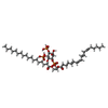  Mass: 1047.088 Da / Num. of mol.: 1 / Source method: obtained synthetically / Formula: C47H85O19P3 / Feature type: SUBJECT OF INVESTIGATION Mass: 1047.088 Da / Num. of mol.: 1 / Source method: obtained synthetically / Formula: C47H85O19P3 / Feature type: SUBJECT OF INVESTIGATION |
| Has ligand of interest | Y |
| Has protein modification | N |
-Experimental details
-Experiment
| Experiment | Method: ELECTRON MICROSCOPY |
|---|---|
| EM experiment | Aggregation state: PARTICLE / 3D reconstruction method: single particle reconstruction |
- Sample preparation
| Component | Name: mouse PMCA captured in E1-ATP in the presence of Calcium Type: COMPLEX Details: prepared as separate cDNAs for transient (co)transfection of tsA201 nptn/basi double KO cells Entity ID: #1 / Source: RECOMBINANT |
|---|---|
| Molecular weight | Value: 0.18 MDa / Experimental value: NO |
| Source (natural) | Organism: |
| Source (recombinant) | Organism: Homo sapiens (human) |
| Buffer solution | pH: 7.4 / Details: Tris 20mM NaCl 150mM |
| Specimen | Conc.: 2 mg/ml / Embedding applied: NO / Shadowing applied: NO / Staining applied: NO / Vitrification applied: YES / Details: The complex was monodisperse |
| Specimen support | Grid material: COPPER / Grid type: Quantifoil R1.2/1.3 |
| Vitrification | Instrument: FEI VITROBOT MARK IV / Cryogen name: ETHANE / Humidity: 95 % / Chamber temperature: 277 K |
- Electron microscopy imaging
| Experimental equipment |  Model: Titan Krios / Image courtesy: FEI Company |
|---|---|
| Microscopy | Model: TFS KRIOS |
| Electron gun | Electron source:  FIELD EMISSION GUN / Accelerating voltage: 300 kV / Illumination mode: FLOOD BEAM FIELD EMISSION GUN / Accelerating voltage: 300 kV / Illumination mode: FLOOD BEAM |
| Electron lens | Mode: BRIGHT FIELD / Nominal magnification: 55000 X / Calibrated magnification: 105000 X / Nominal defocus max: 2500 nm / Nominal defocus min: 1100 nm / Cs: 2.7 mm / C2 aperture diameter: 50 µm / Alignment procedure: ZEMLIN TABLEAU |
| Specimen holder | Cryogen: NITROGEN / Specimen holder model: FEI TITAN KRIOS AUTOGRID HOLDER / Temperature (max): 113 K / Temperature (min): 93 K |
| Image recording | Average exposure time: 3.5 sec. / Electron dose: 57 e/Å2 / Film or detector model: GATAN K3 (6k x 4k) / Num. of real images: 8076 |
| EM imaging optics | Energyfilter name: GIF Bioquantum / Energyfilter slit width: 20 eV |
- Processing
| EM software |
| ||||||||||||||||||||||||||||||||||||||||||||||||||
|---|---|---|---|---|---|---|---|---|---|---|---|---|---|---|---|---|---|---|---|---|---|---|---|---|---|---|---|---|---|---|---|---|---|---|---|---|---|---|---|---|---|---|---|---|---|---|---|---|---|---|---|
| CTF correction | Type: PHASE FLIPPING AND AMPLITUDE CORRECTION | ||||||||||||||||||||||||||||||||||||||||||||||||||
| Symmetry | Point symmetry: C1 (asymmetric) | ||||||||||||||||||||||||||||||||||||||||||||||||||
| 3D reconstruction | Resolution: 3.39 Å / Resolution method: FSC 0.143 CUT-OFF / Num. of particles: 300272 / Algorithm: FOURIER SPACE / Num. of class averages: 1 / Symmetry type: POINT | ||||||||||||||||||||||||||||||||||||||||||||||||||
| Atomic model building | B value: 161.4 / Protocol: OTHER / Space: REAL | ||||||||||||||||||||||||||||||||||||||||||||||||||
| Atomic model building | PDB-ID: 6A69 Accession code: 6A69 / Source name: PDB / Type: experimental model | ||||||||||||||||||||||||||||||||||||||||||||||||||
| Refinement | Highest resolution: 3.39 Å Stereochemistry target values: REAL-SPACE (WEIGHTED MAP SUM AT ATOM CENTERS) | ||||||||||||||||||||||||||||||||||||||||||||||||||
| Refine LS restraints |
|

