 Movie
Movie Controller

[English] 日本語
 Yorodumi
- PDB-9gne: CryoEM structure of mammalian AAP in complex with acetyl-alanyl-c... -
+ Open data

- Basic information
| Entry | Database: PDB / ID: 9gne | ||||||||||||
|---|---|---|---|---|---|---|---|---|---|---|---|---|---|
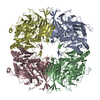
| Title | CryoEM structure of mammalian AAP in complex with acetyl-alanyl-chloromethylketone | ||||||||||||
 Components Components | Acylamino-acid-releasing enzyme | ||||||||||||
 Keywords Keywords | HYDROLASE / covalent inhibitor / serine-protease / flexible active site / oligopeptidase / self-assembly | ||||||||||||
| Function / homology |  Function and homology information Function and homology informationacylaminoacyl-peptidase / omega peptidase activity / serine-type endopeptidase activity / proteolysis / cytoplasm Similarity search - Function | ||||||||||||
| Biological species |  | ||||||||||||
| Method | ELECTRON MICROSCOPY / single particle reconstruction / cryo EM / Resolution: 3.2 Å | ||||||||||||
 Authors Authors | Kiss-Szeman, A.J. / Jakli, I. / Hosogi, N. / Banoczi, Z. / Harmat, V. / Memyhard, D.K. / Perczel, A. | ||||||||||||
| Funding support |  Hungary, European Union, 3items Hungary, European Union, 3items
| ||||||||||||
 Citation Citation | Journal: Protein Sci / Year: 2025 Title: Ligand binding Pro-miscuity of acylpeptide hydrolase, structural analysis of a detoxifying serine hydrolase. Authors: Anna J Kiss-Szemán / Luca Takács / Imre Jákli / Zoltán Bánóczi / Naoki Hosogi / Daouda A K Traore / Veronika Harmat / András Perczel / Dóra K Menyhárd /   Abstract: Acylpeptide hydrolase (APEH) or acylaminoacyl-peptidase (AAP) is a serine hydrolase that regulates protein metabolism. It can also bind to and process unusual substrates, acting as a detoxifier. To ...Acylpeptide hydrolase (APEH) or acylaminoacyl-peptidase (AAP) is a serine hydrolase that regulates protein metabolism. It can also bind to and process unusual substrates, acting as a detoxifier. To better understand its promiscuous specificity, we determined the cryo-EM structures of mammalian APEH complexed with classical serine protease partners: a chloromethyl-ketone (CMK) inhibitor, an organophosphate (OP) pesticide (dichlorvos), and benzenesulfonyl-fluoride. Since CMK derivatives of N-acetylated peptides were suggested to induce apoptosis by inhibiting APEH, while OP complexes may serve as biomarkers of OP exposure and are linked to cognitive enhancement, these complexes carry physiological significance. We identified a unique strand-breaker Pro residue in the hydrolase domain, which relaxes the active site into a partially inactivated but more spacious conformation, transforming the classical serine protease apparatus into a versatile yet potent hydrolysis center with broad specificity, distinguishing the mammalian enzyme not only from other APEHs but also from serine α/β hydrolases sharing essentially the same fold. | ||||||||||||
| History |
|
- Structure visualization
| Structure viewer | Molecule: MolmilJmol/JSmol |
|---|
- Downloads & links
-Download
| PDBx/mmCIF format | 9gne.cif.gz | 491.4 KB | Display | PDBx/mmCIF format |
|---|---|---|---|---|
| PDB format | pdb9gne.ent.gz | Display | PDB format | |
| PDBx/mmJSON format | 9gne.json.gz | Tree view | PDBx/mmJSON format | |
| Others |  Other downloads Other downloads |
-Validation report
| Arichive directory | https://data.pdbj.org/pub/pdb/validation_reports/gn/9gneftp://data.pdbj.org/pub/pdb/validation_reports/gn/9gne | HTTPS FTP |
|---|
-Related structure data
| Related structure data |  51464MC  9gouC  9hxqC  9s6bC M: map data used to model this data C: citing same article ( |
|---|---|
| Similar structure data |


-Links
 PDBj
PDBj


- Assembly
| Deposited unit | 
|
|---|---|
| 1 |
|
-Components
| #1: Protein | Mass: 81324.391 Da / Num. of mol.: 4 / Source method: isolated from a natural source / Source: (natural) #2: Chemical | ChemComp-A1INA / ~{ Mass: 131.173 Da / Num. of mol.: 4 / Source method: obtained synthetically / Formula: C6H13NO2 / Feature type: SUBJECT OF INVESTIGATION Has ligand of interest | Y | Has protein modification | N | |
|---|
-Experimental details
-Experiment
| Experiment | Method: ELECTRON MICROSCOPY |
|---|---|
| EM experiment | Aggregation state: PARTICLE / 3D reconstruction method: single particle reconstruction |
- Sample preparation
| Component | Name: homotetramer of acylaminoacyl-peptidase (isolated from porcine liver) in covalent komplex with acetyl-alanyl-chloromethylketone Type: ORGANELLE OR CELLULAR COMPONENT / Entity ID: #1 / Source: NATURAL |
|---|---|
| Molecular weight | Experimental value: NO |
| Source (natural) | Organism: |
| Buffer solution | pH: 7.5 |
| Buffer component | Conc.: 10 mM / Name: TRIS / Formula: TRIS |
| Specimen | Conc.: 0.5 mg/ml / Embedding applied: NO / Shadowing applied: NO / Staining applied: NO / Vitrification applied: YES |
| Specimen support | Grid material: COPPER / Grid mesh size: 400 divisions/in. / Grid type: Quantifoil R1.2/1.3 |
| Vitrification | Instrument: LEICA EM GP / Cryogen name: ETHANE |
- Electron microscopy imaging
| Microscopy | Model: JEOL CRYO ARM 300 |
|---|---|
| Electron gun | Electron source:  FIELD EMISSION GUN / Accelerating voltage: 300 kV / Illumination mode: FLOOD BEAM FIELD EMISSION GUN / Accelerating voltage: 300 kV / Illumination mode: FLOOD BEAM |
| Electron lens | Mode: BRIGHT FIELD / Nominal magnification: 80000 X / Nominal defocus max: 2500 nm / Nominal defocus min: 500 nm |
| Specimen holder | Cryogen: NITROGEN |
| Image recording | Electron dose: 40 e/Å2 / Film or detector model: GATAN K3 (6k x 4k) |
- Processing
| EM software |
| ||||||||||||||||||||||||
|---|---|---|---|---|---|---|---|---|---|---|---|---|---|---|---|---|---|---|---|---|---|---|---|---|---|
| CTF correction | Type: PHASE FLIPPING AND AMPLITUDE CORRECTION | ||||||||||||||||||||||||
| Particle selection | Num. of particles selected: 2151591 | ||||||||||||||||||||||||
| Symmetry | Point symmetry: D2 (2x2 fold dihedral) | ||||||||||||||||||||||||
| 3D reconstruction | Resolution: 3.2 Å / Resolution method: FSC 0.143 CUT-OFF / Num. of particles: 412960 / Num. of class averages: 7 / Symmetry type: POINT | ||||||||||||||||||||||||
| Refine LS restraints |
|
