 Movie
Movie Controller

[English] 日本語
 Yorodumi
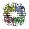
- EMDB-51501: Cryo-EM structure of acylaminoacyl-peptidase in complex with dich... -
+ Open data

- Basic information
| Entry |  | ||||||||||||
|---|---|---|---|---|---|---|---|---|---|---|---|---|---|
| Title | Cryo-EM structure of acylaminoacyl-peptidase in complex with dichlorvos | ||||||||||||
 Map data Map data | |||||||||||||
 Sample Sample |
| ||||||||||||
 Keywords Keywords | serine-protease / covalent inhibitor / dichlorvos / dimethoxy-phosphate / acyl-peptide-hydrolase / oxidized protein hydrolase / APEH / AARE / AAP / OPH / HYDROLASE | ||||||||||||
| Function / homology |  Function and homology information Function and homology informationacylaminoacyl-peptidase / omega peptidase activity / serine-type endopeptidase activity / proteolysis / cytoplasm Similarity search - Function | ||||||||||||
| Biological species |  | ||||||||||||
| Method | single particle reconstruction / cryo EM / Resolution: 2.65 Å | ||||||||||||
 Authors Authors | Kiss-Szeman AJ / Traore D / Jakli I / Harmat V / Menyhard DK / Perczel A | ||||||||||||
| Funding support |  Hungary, European Union, 3 items Hungary, European Union, 3 items
| ||||||||||||
 Citation Citation | Journal: Protein Sci / Year: 2025 Title: Ligand binding Pro-miscuity of acylpeptide hydrolase, structural analysis of a detoxifying serine hydrolase. Authors: Anna J Kiss-Szemán / Luca Takács / Imre Jákli / Zoltán Bánóczi / Naoki Hosogi / Daouda A K Traore / Veronika Harmat / András Perczel / Dóra K Menyhárd /   Abstract: Acylpeptide hydrolase (APEH) or acylaminoacyl-peptidase (AAP) is a serine hydrolase that regulates protein metabolism. It can also bind to and process unusual substrates, acting as a detoxifier. To ...Acylpeptide hydrolase (APEH) or acylaminoacyl-peptidase (AAP) is a serine hydrolase that regulates protein metabolism. It can also bind to and process unusual substrates, acting as a detoxifier. To better understand its promiscuous specificity, we determined the cryo-EM structures of mammalian APEH complexed with classical serine protease partners: a chloromethyl-ketone (CMK) inhibitor, an organophosphate (OP) pesticide (dichlorvos), and benzenesulfonyl-fluoride. Since CMK derivatives of N-acetylated peptides were suggested to induce apoptosis by inhibiting APEH, while OP complexes may serve as biomarkers of OP exposure and are linked to cognitive enhancement, these complexes carry physiological significance. We identified a unique strand-breaker Pro residue in the hydrolase domain, which relaxes the active site into a partially inactivated but more spacious conformation, transforming the classical serine protease apparatus into a versatile yet potent hydrolysis center with broad specificity, distinguishing the mammalian enzyme not only from other APEHs but also from serine α/β hydrolases sharing essentially the same fold. | ||||||||||||
| History |
|
- Structure visualization
| Supplemental images |
|---|

- Downloads & links
-EMDB archive
| Map data | emd_51501.map.gz | 206.2 MB | EMDB map data format | |
|---|---|---|---|---|
| Header (meta data) | emd-51501-v30.xmlemd-51501.xml | 18.6 KB 18.6 KB | Display Display | EMDB header |
| FSC (resolution estimation) | emd_51501_fsc.xml | 13.1 KB | Display | FSC data file |
| Images |  emd_51501.png emd_51501.png | 44.8 KB | ||
| Filedesc metadata | emd-51501.cif.gz | 6.2 KB | ||
| Others | emd_51501_half_map_1.map.gzemd_51501_half_map_2.map.gz | 225.9 MB 225.9 MB | ||
| Archive directory |  http://ftp.pdbj.org/pub/emdb/structures/EMD-51501ftp://ftp.pdbj.org/pub/emdb/structures/EMD-51501 http://ftp.pdbj.org/pub/emdb/structures/EMD-51501ftp://ftp.pdbj.org/pub/emdb/structures/EMD-51501 | HTTPS FTP |
-Related structure data
| Related structure data |  9gouMC  9gneC  9hxqC  9s6bC M: atomic model generated by this map C: citing same article ( |
|---|---|
| Similar structure data |


-Links
| EMDB pages | EMDB (EBI/PDBe) / EMDataResource |
|---|---|
| Related items in Molecule of the Month |


-Map
| File | Download / File: emd_51501.map.gz / Format: CCP4 / Size: 244.1 MB / Type: IMAGE STORED AS FLOATING POINT NUMBER (4 BYTES) | ||||||||||||||||||||||||||||||||||||
|---|---|---|---|---|---|---|---|---|---|---|---|---|---|---|---|---|---|---|---|---|---|---|---|---|---|---|---|---|---|---|---|---|---|---|---|---|---|
| Projections & slices | Image control
Images are generated by Spider. | ||||||||||||||||||||||||||||||||||||
| Voxel size | X=Y=Z: 0.7519 Å | ||||||||||||||||||||||||||||||||||||
| Density |
| ||||||||||||||||||||||||||||||||||||
| Symmetry | Space group: 1 | ||||||||||||||||||||||||||||||||||||
| Details | EMDB XML:
|
 Z (Sec.)
 Y (Row.)
 X (Col.)


-Supplemental data
-Half map: #2
| File | emd_51501_half_map_1.map | ||||||||||||
|---|---|---|---|---|---|---|---|---|---|---|---|---|---|
| Projections & Slices |
| ||||||||||||
| Density Histograms |


-Half map: #1
| File | emd_51501_half_map_2.map | ||||||||||||
|---|---|---|---|---|---|---|---|---|---|---|---|---|---|
| Projections & Slices |
| ||||||||||||
| Density Histograms |


- Sample components
-Entire : homotetramer of AAP
| Entire | Name: homotetramer of AAP |
|---|---|
| Components |
|
-Supramolecule #1: homotetramer of AAP
| Supramolecule | Name: homotetramer of AAP / type: organelle_or_cellular_component / ID: 1 / Parent: 0 / Macromolecule list: #1 |
|---|---|
| Source (natural) | Organism: |
-Macromolecule #1: Acylamino-acid-releasing enzyme
| Macromolecule | Name: Acylamino-acid-releasing enzyme / type: protein_or_peptide / ID: 1 / Number of copies: 4 / Enantiomer: LEVO / EC number: acylaminoacyl-peptidase |
|---|---|
| Source (natural) | Organism: |
| Molecular weight | Theoretical: 81.324391 KDa |
| Sequence | String: MERQVLLSEP EEAAALYRGL SRQPALSAAC LGPEVTTQYG GRYRTVHTEW TQRDLERMEN IRFCRQYLVF HDGDSVVFAG PAGNSVETR GELLSRESPS GTMKAVLRKA GGTGTAEEKQ FLEVWEKNRK LKSFNLSALE KHGPVYEDDC FGCLSWSHSE T HLLYVADK ...String: MERQVLLSEP EEAAALYRGL SRQPALSAAC LGPEVTTQYG GRYRTVHTEW TQRDLERMEN IRFCRQYLVF HDGDSVVFAG PAGNSVETR GELLSRESPS GTMKAVLRKA GGTGTAEEKQ FLEVWEKNRK LKSFNLSALE KHGPVYEDDC FGCLSWSHSE T HLLYVADK KRPKAESFFQ TKALDVTGSD DEMARTKKPD QAIKGDQFLF YEDWGENMVS KSTPVLCVLD IESGNISVLE GV PESVSPG QAFWAPGDTG VVFVGWWHEP FRLGIRFCTN RRSALYYVDL TGGKCELLSD ESVAVTSPRL SPDQCRIVYL RFP SLVPHQ QCGQLCLYDW YTRVTSVVVD IVPRQLGEDF SGIYCSLLPL GCWSADSQRV VFDSPQRSRQ DLFAVDTQMG SVTS LTAGG SGGSWKLLTI DRDLMVVQFS TPSVPPSLKV GFLPPAGKEQ AVSWVSLEEA EPFPDISWSI RVLQPPPQQE HVQYA GLDF EAILLQPSNS PEKTQVPMVV MPHGGPHSSF VTAWMLFPAM LCKMGFAVLL VNYRGSTGFG QDSILSLPGN VGHQDV KDV QFAVEQVLQE EHFDAGRVAL MGGSHGGFLS CHLIGQYPET YSACVVRNPV INIASMMGST DIPDWCMVEA GFSYSSD CL PDLSVWAAML DKSPIKYAPQ VKTPLLLMLG QEDRRVPFKQ GMEYYRVLKA RNVPVRLLLY PKSTHALSEV EVESDSFM N AVLWLCTHLG S UniProtKB: Acylamino-acid-releasing enzyme |
-Macromolecule #2: dimethyl hydrogen phosphite
| Macromolecule | Name: dimethyl hydrogen phosphite / type: ligand / ID: 2 / Number of copies: 4 / Formula: A1ING |
|---|---|
| Molecular weight | Theoretical: 110.049 Da |
-Experimental details
-Structure determination
| Method | cryo EM |
|---|---|
 Processing Processing | single particle reconstruction |
| Aggregation state | particle |
-Sample preparation
| Concentration | 0.5 mg/mL |
|---|---|
| Buffer | pH: 7.5 / Component - Concentration: 10.0 mM / Component - Formula: TRIS / Component - Name: TRIS |
| Grid | Model: Quantifoil R2/2 / Material: COPPER / Mesh: 300 / Pretreatment - Type: GLOW DISCHARGE |
| Vitrification | Cryogen name: ETHANE / Chamber humidity: 95 % / Chamber temperature: 277 K / Instrument: FEI VITROBOT MARK IV |
- Electron microscopy
| Microscope | TFS KRIOS |
|---|---|
| Image recording | Film or detector model: TFS FALCON 4i (4k x 4k) / Number grids imaged: 2 / Number real images: 12467 / Average exposure time: 1.9 sec. / Average electron dose: 11.9 e/Å2 |
| Electron beam | Acceleration voltage: 300 kV / Electron source:  FIELD EMISSION GUN FIELD EMISSION GUN |
| Electron optics | C2 aperture diameter: 50.0 µm / Illumination mode: FLOOD BEAM / Imaging mode: OTHER / Cs: 2.7 mm / Nominal defocus max: 1.5 µm / Nominal defocus min: 0.5 µm / Nominal magnification: 165000 |
| Sample stage | Cooling holder cryogen: NITROGEN |
| Experimental equipment |  Model: Titan Krios / Image courtesy: FEI Company |
+Image processing

-Atomic model buiding 1
| Refinement | Protocol: RIGID BODY FIT |
|---|---|
| Output model | PDB-9gou: |
