National Research Development and Innovation Office (NKFIH)
Thematic Excellence Program 2019, Synth+
Hungary
National Research Development and Innovation Office (NKFIH)
K116305
Hungary
Citation
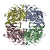
Journal: Chem Sci / Year: 2022 Title: A carbapenem antibiotic inhibiting a mammalian serine protease: structure of the acylaminoacyl peptidase-meropenem complex. Authors: Anna J Kiss-Szemán / Luca Takács / Zoltán Orgován / Pál Stráner / Imre Jákli / Gitta Schlosser / Simonas Masiulis / Veronika Harmat / Dóra K Menyhárd / András Perczel / Abstract: The structure of porcine AAP (pAAP) in a covalently bound complex with meropenem was determined by cryo-EM to 2.1 Å resolution, showing the mammalian serine-protease inhibited by a carbapenem ...The structure of porcine AAP (pAAP) in a covalently bound complex with meropenem was determined by cryo-EM to 2.1 Å resolution, showing the mammalian serine-protease inhibited by a carbapenem antibiotic. AAP is a modulator of the ubiquitin-proteasome degradation system and the site of a drug-drug interaction between the widely used antipsychotic, valproate and carbapenems. The active form of pAAP - a toroidal tetramer - binds four meropenem molecules covalently linked to the catalytic Ser587 of the serine-protease triad, in an acyl-enzyme state. AAP is hindered from fully processing the antibiotic by the displacement and protonation of His707 of the catalytic triad. We show that AAP is made susceptible to the association by its unusually sheltered active pockets and flexible catalytic triads, while the carbapenems possess sufficiently small substituents on their β-lactam rings to fit into the shallow substrate-specificity pocket of the enzyme.
History
Deposition
Jan 18, 2022
Deposition site: PDBE / Processing site: PDBE
Revision 1.0
Nov 16, 2022
Provider: repository / Type: Initial release
Revision 1.0
Nov 16, 2022
Data content type: EM metadata / Data content type: EM metadata / Provider: repository / Type: Initial release
Revision 1.0
Nov 16, 2022
Data content type: Image / Data content type: Image / Provider: repository / Type: Initial release
Revision 1.0
Nov 16, 2022
Data content type: Primary map / Data content type: Primary map / Provider: repository / Type: Initial release
Revision 1.0
Nov 16, 2022
Data content type: Image / Data content type: Image / Provider: repository / Type: Initial release
Revision 1.0
Nov 16, 2022
Data content type: Primary map / Data content type: Primary map / Provider: repository / Type: Initial release
Revision 1.0
Nov 16, 2022
Data content type: Image / Data content type: Image / Provider: repository / Type: Initial release
Revision 1.0
Nov 16, 2022
Data content type: Primary map / Data content type: Primary map / Provider: repository / Type: Initial release
Data content type: EM metadata / Data content type: EM metadata / EM metadata / Group: Data processing / Experimental summary / Data content type: EM metadata / EM metadata / Category: em_admin / em_software / Data content type: EM metadata / EM metadata / Item: _em_admin.last_update / _em_software.name
Instrument: FEI VITROBOT MARK IV / Cryogen name: ETHANE / Humidity: 95 % / Chamber temperature: 277 K
-
Electron microscopy imaging
Experimental equipment
Model: Titan Krios / Image courtesy: FEI Company
Microscopy
Model: FEI TITAN KRIOS
Electron gun
Electron source: FIELD EMISSION GUN / Accelerating voltage: 300 kV / Illumination mode: FLOOD BEAM
Electron lens
Mode: BRIGHT FIELD / Nominal magnification: 130000 X / Nominal defocus max: 3000 nm / Nominal defocus min: 1000 nm
Image recording
Average exposure time: 5.93 sec. / Electron dose: 41 e/Å2 / Film or detector model: FEI FALCON IV (4k x 4k) / Num. of grids imaged: 4 / Num. of real images: 11625
In the structure databanks used in Yorodumi, some data are registered as the other names, "COVID-19 virus" and "2019-nCoV". Here are the details of the virus and the list of structure data.
Jan 31, 2019. EMDB accession codes are about to change! (news from PDBe EMDB page)
EMDB accession codes are about to change! (news from PDBe EMDB page)
The allocation of 4 digits for EMDB accession codes will soon come to an end. Whilst these codes will remain in use, new EMDB accession codes will include an additional digit and will expand incrementally as the available range of codes is exhausted. The current 4-digit format prefixed with “EMD-” (i.e. EMD-XXXX) will advance to a 5-digit format (i.e. EMD-XXXXX), and so on. It is currently estimated that the 4-digit codes will be depleted around Spring 2019, at which point the 5-digit format will come into force.
The EM Navigator/Yorodumi systems omit the EMD- prefix.
Related info.:Q: What is EMD? / ID/Accession-code notation in Yorodumi/EM Navigator
Yorodumi is a browser for structure data from EMDB, PDB, SASBDB, etc.
This page is also the successor to EM Navigator detail page, and also detail information page/front-end page for Omokage search.
The word "yorodu" (or yorozu) is an old Japanese word meaning "ten thousand". "mi" (miru) is to see.
Related info.:EMDB / PDB / SASBDB / Comparison of 3 databanks / Yorodumi Search / Aug 31, 2016. New EM Navigator & Yorodumi / Yorodumi Papers / Jmol/JSmol / Function and homology information / Changes in new EM Navigator and Yorodumi
 Movie
Movie Controller


 Open data
 Basic information
 Components
 Keywords
 Function and homology information
 Authors
 Hungary, 5items
  Citation
 Structure visualization
 Downloads & links
 Other downloads
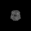


 PDBj
PDBj


 Assembly
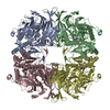
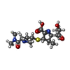
 Mass: 385.478 Da / Num. of mol.: 4 / Source method: obtained synthetically / Formula: C17H27N3O5S / Feature type: SUBJECT OF INVESTIGATION / Comment: antibiotic*YM
 Sample preparation
 Electron microscopy imaging
 FIELD EMISSION GUN / Accelerating voltage: 300 kV / Illumination mode: FLOOD BEAM
 Processing