[English] 日本語
 Yorodumi
- PDB-9gkx: Crystal Structure of Rhizorhabdus wittichii Dimethoate hydrolase ... -
+ Open data
- Basic information
| Entry | Database: PDB / ID: 9gkx | |||||||||
|---|---|---|---|---|---|---|---|---|---|---|
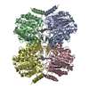
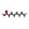
| Title | Crystal Structure of Rhizorhabdus wittichii Dimethoate hydrolase (DmhA) in complex with SAHA | |||||||||
 Components Components | Dimethoate hydrolase | |||||||||
 Keywords Keywords | HYDROLASE / Inhibitor / SAHA / Deacetylase / Deacylase | |||||||||
| Function / homology |  Function and homology information Function and homology informationhistone deacetylase activity / epigenetic regulation of gene expression / hydrolase activity / metal ion binding Similarity search - Function | |||||||||
| Biological species |  Rhizorhabdus wittichii DC-6 (bacteria) Rhizorhabdus wittichii DC-6 (bacteria) | |||||||||
| Method |  X-RAY DIFFRACTION / X-RAY DIFFRACTION /  SYNCHROTRON / SYNCHROTRON /  MOLECULAR REPLACEMENT / Resolution: 1.75 Å MOLECULAR REPLACEMENT / Resolution: 1.75 Å | |||||||||
 Authors Authors | Graf, L.G. / Lammers, M. / Schulze, S. / Palm, G.J. | |||||||||
| Funding support |  Germany, 2items Germany, 2items
| |||||||||
 Citation Citation |  Journal: Nat Commun / Year: 2024 Journal: Nat Commun / Year: 2024Title: Distribution and diversity of classical deacylases in bacteria. Authors: Graf, L.G. / Moreno-Yruela, C. / Qin, C. / Schulze, S. / Palm, G.J. / Schmoker, O. / Wang, N. / Hocking, D.M. / Jebeli, L. / Girbardt, B. / Berndt, L. / Dorre, B. / Weis, D.M. / Janetzky, M. ...Authors: Graf, L.G. / Moreno-Yruela, C. / Qin, C. / Schulze, S. / Palm, G.J. / Schmoker, O. / Wang, N. / Hocking, D.M. / Jebeli, L. / Girbardt, B. / Berndt, L. / Dorre, B. / Weis, D.M. / Janetzky, M. / Albrecht, D. / Zuhlke, D. / Sievers, S. / Strugnell, R.A. / Olsen, C.A. / Hofmann, K. / Lammers, M. | |||||||||
| History |
|
- Structure visualization
| Structure viewer | Molecule:  Molmil Molmil Jmol/JSmol Jmol/JSmol |
|---|
- Downloads & links
- Download
| PDBx/mmCIF format |  9gkx.cif.gz 9gkx.cif.gz | 413.3 KB | Display |  PDBx/mmCIF format PDBx/mmCIF format |
|---|---|---|---|---|
| PDB format |  pdb9gkx.ent.gz pdb9gkx.ent.gz | 261.4 KB | Display |  PDB format PDB format |
| PDBx/mmJSON format |  9gkx.json.gz 9gkx.json.gz | Tree view |  PDBx/mmJSON format PDBx/mmJSON format | |
| Others |  Other downloads Other downloads |
-Validation report
| Summary document |  9gkx_validation.pdf.gz 9gkx_validation.pdf.gz | 2.9 MB | Display |  wwPDB validaton report wwPDB validaton report |
|---|---|---|---|---|
| Full document |  9gkx_full_validation.pdf.gz 9gkx_full_validation.pdf.gz | 2.9 MB | Display | |
| Data in XML |  9gkx_validation.xml.gz 9gkx_validation.xml.gz | 81.4 KB | Display | |
| Data in CIF |  9gkx_validation.cif.gz 9gkx_validation.cif.gz | 115.4 KB | Display | |
| Arichive directory |  https://data.pdbj.org/pub/pdb/validation_reports/gk/9gkx https://data.pdbj.org/pub/pdb/validation_reports/gk/9gkx ftp://data.pdbj.org/pub/pdb/validation_reports/gk/9gkx ftp://data.pdbj.org/pub/pdb/validation_reports/gk/9gkx | HTTPS FTP |
-Related structure data
| Related structure data |  9gkuC  9gkvC  9gkwC  9gkyC  9gkzC  9gl0C  9gl1C  9glbC  9gn1C  9gn6C  9gn7C C: citing same article ( |
|---|---|
| Similar structure data | Similarity search - Function & homology  F&H Search F&H Search |
- Links
Links
- Assembly
| Deposited unit | 
| ||||||||
|---|---|---|---|---|---|---|---|---|---|
| 1 |
| ||||||||
| Unit cell |
|
- Components
-Protein , 1 types, 4 molecules ABCD
| #1: Protein | Mass: 40816.195 Da / Num. of mol.: 4 Source method: isolated from a genetically manipulated source Details: His6x-Tag fusion-protein / Source: (gene. exp.)  Rhizorhabdus wittichii DC-6 (bacteria) / Strain: DC-6 / KACC 16600 / Gene: dmhA / Plasmid: pET-45b(+) / Production host: Rhizorhabdus wittichii DC-6 (bacteria) / Strain: DC-6 / KACC 16600 / Gene: dmhA / Plasmid: pET-45b(+) / Production host:  |
|---|
-Non-polymers , 5 types, 1792 molecules 


| #2: Chemical | | #3: Chemical | ChemComp-ZN / #4: Chemical | ChemComp-K / #5: Chemical | #6: Water | ChemComp-HOH / | |
|---|
-Details
| Has ligand of interest | Y |
|---|---|
| Has protein modification | N |
-Experimental details
-Experiment
| Experiment | Method:  X-RAY DIFFRACTION / Number of used crystals: 1 X-RAY DIFFRACTION / Number of used crystals: 1 |
|---|
- Sample preparation
| Crystal | Density Matthews: 2.85 Å3/Da / Density % sol: 56.86 % |
|---|---|
| Crystal grow | Temperature: 293.15 K / Method: vapor diffusion, sitting drop / pH: 8 / Details: 0.2 M Potassium fluoride pH 7.3 20% (w/v) PEG3350 |
-Data collection
| Diffraction | Mean temperature: 100 K / Serial crystal experiment: N |
|---|---|
| Diffraction source | Source:  SYNCHROTRON / Site: SYNCHROTRON / Site:  BESSY BESSY  / Beamline: 14.1 / Wavelength: 0.9184 Å / Beamline: 14.1 / Wavelength: 0.9184 Å |
| Detector | Type: DECTRIS PILATUS3 S 6M / Detector: PIXEL / Date: Apr 4, 2024 |
| Radiation | Protocol: SINGLE WAVELENGTH / Monochromatic (M) / Laue (L): M / Scattering type: x-ray |
| Radiation wavelength | Wavelength: 0.9184 Å / Relative weight: 1 |
| Reflection | Resolution: 1.75→50 Å / Num. obs: 174428 / % possible obs: 99.7 % / Observed criterion σ(I): -3 / Redundancy: 6.9 % / CC1/2: 0.994 / Rrim(I) all: 0.229 / Rsym value: 0.21 / Χ2: 0.99 / Net I/σ(I): 7.68 |
| Reflection shell | Resolution: 1.75→1.86 Å / Num. unique obs: 28058 / CC1/2: 0.488 / Χ2: 0.9 / % possible all: 99.6 |
- Processing
| Software |
| ||||||||||||||||
|---|---|---|---|---|---|---|---|---|---|---|---|---|---|---|---|---|---|
| Refinement | Method to determine structure:  MOLECULAR REPLACEMENT / Resolution: 1.75→46.31 Å / Cross valid method: FREE R-VALUE MOLECULAR REPLACEMENT / Resolution: 1.75→46.31 Å / Cross valid method: FREE R-VALUE
| ||||||||||||||||
| Refinement step | Cycle: LAST / Resolution: 1.75→46.31 Å
|
 Movie
Movie Controller


 PDBj
PDBj


