 Movie
Movie Controller

[English] 日本語
 Yorodumi
- PDB-9g9n: Lipid III flippase WzxE with NB10 and NB7 nanobodies in inward-fa... -
+ Open data

- Basic information
| Entry | Database: PDB / ID: 9g9n | ||||||
|---|---|---|---|---|---|---|---|
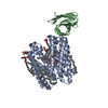
| Title | Lipid III flippase WzxE with NB10 and NB7 nanobodies in inward-facing conformation - crystal 1 | ||||||
 Components Components |
| ||||||
 Keywords Keywords | TRANSPORT PROTEIN / membrane protein / flippase / cell wall / enterobacterial common antigen / lipid III | ||||||
| Function / homology | intramembrane lipid carrier activity / Lipid III flippase WzxE, Proteobacteria / Lipid III flippase WzxE / Polysaccharide biosynthesis protein / : / Polysaccharide biosynthesis protein / enterobacterial common antigen biosynthetic process / plasma membrane / Lipid III flippase Function and homology information Function and homology information | ||||||
| Biological species |   | ||||||
| Method |  X-RAY DIFFRACTION / SYNCHROTRON / MOLECULAR REPLACEMENT / Resolution: 2.8 Å X-RAY DIFFRACTION / SYNCHROTRON / MOLECULAR REPLACEMENT / Resolution: 2.8 Å | ||||||
 Authors Authors | Le Bas, A. / Naismith, J.H. | ||||||
| Funding support |  United Kingdom, 1items United Kingdom, 1items
| ||||||
 Citation Citation | Journal: Open Biology / Year: 2025 Title: Structure of WzxE the lipid III flippase for Enterobacterial Common Antigen polysaccharide. Authors: Le Bas, A. / Clarke, B.R. / Teelucksingh, T. / Lee, M. / El Omari, K. / Giltrap, A.M. / McMahon, S.A. / Liu, H. / Beale, J.H. / Mykhaylyk, V. / Duman, R. / Paterson, N.G. / Ward, P.N. / ...Authors: Le Bas, A. / Clarke, B.R. / Teelucksingh, T. / Lee, M. / El Omari, K. / Giltrap, A.M. / McMahon, S.A. / Liu, H. / Beale, J.H. / Mykhaylyk, V. / Duman, R. / Paterson, N.G. / Ward, P.N. / Harrison, P.J. / Weckener, M. / Pardon, E. / Steyaert, J. / Liu, H. / Quigley, A. / Davis, B.G. / Wagner, A. / Whitfield, C. / Naismith, J.H. | ||||||
| History |
|
- Structure visualization
| Structure viewer | Molecule: MolmilJmol/JSmol |
|---|
- Downloads & links
-Download
| PDBx/mmCIF format | 9g9n.cif.gz | 264 KB | Display | PDBx/mmCIF format |
|---|---|---|---|---|
| PDB format | pdb9g9n.ent.gz | 215.1 KB | Display | PDB format |
| PDBx/mmJSON format | 9g9n.json.gz | Tree view | PDBx/mmJSON format | |
| Others |  Other downloads Other downloads |
-Validation report
| Arichive directory | https://data.pdbj.org/pub/pdb/validation_reports/g9/9g9nftp://data.pdbj.org/pub/pdb/validation_reports/g9/9g9n | HTTPS FTP |
|---|
-Related structure data


-Links
 PDBj
PDBj

- Assembly
| Deposited unit | 
| ||||||||
|---|---|---|---|---|---|---|---|---|---|
| 1 |
| ||||||||
| Unit cell |
|
-Components
| #1: Protein | Mass: 46130.621 Da / Num. of mol.: 1 Source method: isolated from a genetically manipulated source Source: (gene. exp.) |
|---|---|
| #2: Antibody | Mass: 15132.753 Da / Num. of mol.: 1 Source method: isolated from a genetically manipulated source Source: (gene. exp.) |
| #3: Antibody | Mass: 15223.880 Da / Num. of mol.: 1 Source method: isolated from a genetically manipulated source Source: (gene. exp.) |
| Has protein modification | Y |
-Experimental details
-Experiment
| Experiment | Method: X-RAY DIFFRACTION / Number of used crystals: 1 |
|---|
- Sample preparation
| Crystal | Density Matthews: 2.68 Å3/Da / Density % sol: 54.09 % |
|---|---|
| Crystal grow | Temperature: 293 K / Method: lipidic cubic phase / pH: 5 Details: tri-sodium citrate, lithium sulphate, sodium chloride, PEG400, E coli polar lipids, monoolein |
-Data collection
| Diffraction | Mean temperature: 100 K / Serial crystal experiment: N |
|---|---|
| Diffraction source | Source: SYNCHROTRON / Site: Diamond / Beamline: I24 / Wavelength: 0.6199 Å |
| Detector | Type: DECTRIS EIGER2 X CdTe 9M / Detector: PIXEL / Date: Jul 7, 2023 |
| Radiation | Protocol: SINGLE WAVELENGTH / Monochromatic (M) / Laue (L): M / Scattering type: x-ray |
| Radiation wavelength | Wavelength: 0.6199 Å / Relative weight: 1 |
| Reflection | Resolution: 2.8→33.8 Å / Num. obs: 20612 / % possible obs: 99.9 % / Redundancy: 6.8 % / CC1/2: 1 / Net I/σ(I): 6.5 |
| Reflection shell | Resolution: 2.8→2.85 Å / Redundancy: 6.9 % / Mean I/σ(I) obs: 1.3 / Num. unique obs: 1002 / CC1/2: 0.7 / % possible all: 100 |
- Processing
| Software |
| ||||||||||||||||||||||||||||||||||||||||||||||||||||||||||||||||||||||||||||||||||||||||||||||||||||
|---|---|---|---|---|---|---|---|---|---|---|---|---|---|---|---|---|---|---|---|---|---|---|---|---|---|---|---|---|---|---|---|---|---|---|---|---|---|---|---|---|---|---|---|---|---|---|---|---|---|---|---|---|---|---|---|---|---|---|---|---|---|---|---|---|---|---|---|---|---|---|---|---|---|---|---|---|---|---|---|---|---|---|---|---|---|---|---|---|---|---|---|---|---|---|---|---|---|---|---|---|---|
| Refinement | Method to determine structure: MOLECULAR REPLACEMENT / Resolution: 2.8→32.4 Å / SU ML: 0.44 / Cross valid method: THROUGHOUT / σ(F): 1.34 / Phase error: 36.78 / Stereochemistry target values: ML
| ||||||||||||||||||||||||||||||||||||||||||||||||||||||||||||||||||||||||||||||||||||||||||||||||||||
| Solvent computation | Shrinkage radii: 0.9 Å / VDW probe radii: 1.1 Å / Solvent model: FLAT BULK SOLVENT MODEL | ||||||||||||||||||||||||||||||||||||||||||||||||||||||||||||||||||||||||||||||||||||||||||||||||||||
| Refinement step | Cycle: LAST / Resolution: 2.8→32.4 Å
| ||||||||||||||||||||||||||||||||||||||||||||||||||||||||||||||||||||||||||||||||||||||||||||||||||||
| Refine LS restraints |
| ||||||||||||||||||||||||||||||||||||||||||||||||||||||||||||||||||||||||||||||||||||||||||||||||||||
| LS refinement shell |
| ||||||||||||||||||||||||||||||||||||||||||||||||||||||||||||||||||||||||||||||||||||||||||||||||||||
| Refinement TLS params. | Method: refined / Refine-ID: X-RAY DIFFRACTION
| ||||||||||||||||||||||||||||||||||||||||||||||||||||||||||||||||||||||||||||||||||||||||||||||||||||
| Refinement TLS group |
|
