 Movie
Movie Controller

[English] 日本語
 Yorodumi
- PDB-9g93: CryoET structure of the in vitro grown Bacillus anthracis Sap S-layer -
+ Open data

- Basic information
| Entry | Database: PDB / ID: 9g93 | |||||||||||||||||||||
|---|---|---|---|---|---|---|---|---|---|---|---|---|---|---|---|---|---|---|---|---|---|---|
| Title | CryoET structure of the in vitro grown Bacillus anthracis Sap S-layer | |||||||||||||||||||||
 Components Components | S-layer protein sap | |||||||||||||||||||||
 Keywords Keywords | STRUCTURAL PROTEIN / S-layer / exoskeleton / surface array | |||||||||||||||||||||
| Function / homology |  Function and homology information Function and homology information | |||||||||||||||||||||
| Biological species | Bacillus anthracis str. '34F2 ' | |||||||||||||||||||||
| Method | ELECTRON MICROSCOPY / subtomogram averaging / cryo EM / Resolution: 7.2 Å | |||||||||||||||||||||
 Authors Authors | Sogues, A. / Leigh, K. / Van der Verren, S. / Kudryashev, M. / Pak, A. / Halingstad, E.V. / Cecil, A.J. / Fioravanti, A. / Remaut, H. | |||||||||||||||||||||
| Funding support |  Belgium, European Union, Belgium, European Union,  Germany, Germany,  United States, 6items United States, 6items
| |||||||||||||||||||||
 Citation Citation | Journal: Proc Natl Acad Sci U S A / Year: 2024 Title: Architecture of the Sap S-layer of revealed by integrative structural biology. Authors: Adrià Sogues / Kendra Leigh / Ethan V Halingstad / Sander E Van der Verren / Adam J Cecil / Antonella Fioravanti / Alexander J Pak / Misha Kudryashev / Han Remaut / Abstract: is a spore-forming gram-positive bacterium responsible for anthrax, an infectious disease with a high mortality rate and a target of concern due to bioterrorism and long-term site contamination. The ... is a spore-forming gram-positive bacterium responsible for anthrax, an infectious disease with a high mortality rate and a target of concern due to bioterrorism and long-term site contamination. The entire surface of vegetative cells in exponential or stationary growth phase is covered in proteinaceous arrays called S-layers, composed of Sap or EA1 protein, respectively. The Sap S-layer represents an important virulence factor and cell envelope support structure whose paracrystalline nature is essential for its function. However, the spatial organization of Sap in its lattice state remains elusive. Here, we employed cryoelectron tomography and subtomogram averaging to obtain a map of the Sap S-layer from tubular polymers that revealed a conformational switch between the postassembly protomers and the previously available X-ray structure of the condensed monomers. To build and validate an atomic model of the lattice within this map, we used a combination of molecular dynamics simulations, X-ray crystallography, cross-linking mass spectrometry, and biophysics in an integrative structural biology approach. The Sap lattice model produced recapitulates a close-to-physiological arrangement, reveals high-resolution details of lattice contacts, and sheds light on the mechanisms underlying the stability of the Sap layer. | |||||||||||||||||||||
| History |
|
- Structure visualization
| Structure viewer | Molecule: MolmilJmol/JSmol |
|---|
- Downloads & links
-Download
| PDBx/mmCIF format | 9g93.cif.gz | 832.9 KB | Display | PDBx/mmCIF format |
|---|---|---|---|---|
| PDB format | pdb9g93.ent.gz | 639.4 KB | Display | PDB format |
| PDBx/mmJSON format | 9g93.json.gz | Tree view | PDBx/mmJSON format | |
| Others |  Other downloads Other downloads |
-Validation report
| Arichive directory | https://data.pdbj.org/pub/pdb/validation_reports/g9/9g93ftp://data.pdbj.org/pub/pdb/validation_reports/g9/9g93 | HTTPS FTP |
|---|
-Related structure data
| Related structure data | 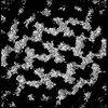 45459MC  8rx2C 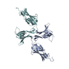 8s80C  8s83C M: map data used to model this data C: citing same article ( |
|---|---|
| Similar structure data |
-Links
 PDBj
PDBj- Assembly
| Deposited unit | 
| ||||||||||||||||||||||||||||||||||||||||||||||||||||||||||||||||||||||||||||||||||||||||||||||
|---|---|---|---|---|---|---|---|---|---|---|---|---|---|---|---|---|---|---|---|---|---|---|---|---|---|---|---|---|---|---|---|---|---|---|---|---|---|---|---|---|---|---|---|---|---|---|---|---|---|---|---|---|---|---|---|---|---|---|---|---|---|---|---|---|---|---|---|---|---|---|---|---|---|---|---|---|---|---|---|---|---|---|---|---|---|---|---|---|---|---|---|---|---|---|---|
| 1 |
| ||||||||||||||||||||||||||||||||||||||||||||||||||||||||||||||||||||||||||||||||||||||||||||||
| Noncrystallographic symmetry (NCS) | NCS domain:
NCS domain segments: Component-ID: 1 / Beg auth comp-ID: LYS / Beg label comp-ID: LYS / End auth comp-ID: GLU / End label comp-ID: GLU / Auth seq-ID: 214 - 808 / Label seq-ID: 213 - 808
NCS ensembles :
NCS oper:
|
-Components
| #1: Protein | Mass: 86734.656 Da / Num. of mol.: 11 Source method: isolated from a genetically manipulated source Source: (gene. exp.)  Strain: 34F2 / Gene: sap, BA_0885, GBAA_0885, BAS0841 / Production host: Has protein modification | N | |
|---|
-Experimental details
-Experiment
| Experiment | Method: ELECTRON MICROSCOPY |
|---|---|
| EM experiment | Aggregation state: 2D ARRAY / 3D reconstruction method: subtomogram averaging |
- Sample preparation
| Component | Name: in vitro S-layer of the Sap assembly domain / Type: COMPLEX Details: 2D S-layer array of a fragment of the Sap protein lacking the SLH cell wall attachment domain, and corresponding to the S-layer assembly domain Entity ID: all / Source: RECOMBINANT |
|---|---|
| Molecular weight | Experimental value: NO |
| Source (natural) | Organism: |
| Source (recombinant) | Organism: |
| Buffer solution | pH: 8 / Details: 10 mM HEPES, pH 8 and 100 mM NaCl |
| Specimen | Embedding applied: NO / Shadowing applied: NO / Staining applied: NO / Vitrification applied: YES |
| Vitrification | Cryogen name: ETHANE |
- Electron microscopy imaging
| Experimental equipment |  Model: Titan Krios / Image courtesy: FEI Company |
|---|---|
| Microscopy | Model: FEI TITAN KRIOS |
| Electron gun | Electron source:  FIELD EMISSION GUN / Accelerating voltage: 300 kV / Illumination mode: FLOOD BEAM FIELD EMISSION GUN / Accelerating voltage: 300 kV / Illumination mode: FLOOD BEAM |
| Electron lens | Mode: BRIGHT FIELD / Nominal magnification: 81000 X / Nominal defocus max: 3500 nm / Nominal defocus min: 2000 nm |
| Image recording | Electron dose: 3.8 e/Å2 / Avg electron dose per subtomogram: 156 e/Å2 / Detector mode: SUPER-RESOLUTION / Film or detector model: GATAN K2 SUMMIT (4k x 4k) / Num. of real images: 1 |
- Processing
| EM software |
| |||||||||||||||||||||||||||||||||||||||||||||||||
|---|---|---|---|---|---|---|---|---|---|---|---|---|---|---|---|---|---|---|---|---|---|---|---|---|---|---|---|---|---|---|---|---|---|---|---|---|---|---|---|---|---|---|---|---|---|---|---|---|---|---|
| CTF correction | Type: PHASE FLIPPING ONLY | |||||||||||||||||||||||||||||||||||||||||||||||||
| Symmetry | Point symmetry: C1 (asymmetric) | |||||||||||||||||||||||||||||||||||||||||||||||||
| 3D reconstruction | Resolution: 7.2 Å / Resolution method: FSC 0.143 CUT-OFF / Num. of particles: 10126 Details: Final half map reconstruction was done in Dynamo and resolution was estimated using RELION 3.1 postprocessing Symmetry type: POINT | |||||||||||||||||||||||||||||||||||||||||||||||||
| EM volume selection | Num. of tomograms: 12 / Num. of volumes extracted: 117502 | |||||||||||||||||||||||||||||||||||||||||||||||||
| Refinement | Cross valid method: NONE Stereochemistry target values: GeoStd + Monomer Library + CDL v1.2 | |||||||||||||||||||||||||||||||||||||||||||||||||
| Displacement parameters | Biso mean: 50 Å2 | |||||||||||||||||||||||||||||||||||||||||||||||||
| Refine LS restraints |
| |||||||||||||||||||||||||||||||||||||||||||||||||
| Refine LS restraints NCS |
|
