 Movie
Movie Controller


+ Open data

- Basic information
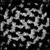
| Entry | Database: PDB / ID: 8s83 | ||||||
|---|---|---|---|---|---|---|---|
| Title | Domains 3 and 6 of Sap S-layer protein from Bacillus anthracis | ||||||
 Components Components | (S-layer protein sap) x 2 | ||||||
 Keywords Keywords | STRUCTURAL PROTEIN / S-layer / Sap / Anthracis / anthrax | ||||||
| Function / homology |  Function and homology information Function and homology information | ||||||
| Biological species |  | ||||||
| Method |  X-RAY DIFFRACTION / SYNCHROTRON / MOLECULAR REPLACEMENT / Resolution: 2.07 Å X-RAY DIFFRACTION / SYNCHROTRON / MOLECULAR REPLACEMENT / Resolution: 2.07 Å | ||||||
 Authors Authors | Sogues, A. / Remaut, H. | ||||||
| Funding support | European Union, 1items
| ||||||
 Citation Citation | Journal: Proc Natl Acad Sci U S A / Year: 2024 Title: Architecture of the Sap S-layer of revealed by integrative structural biology. Authors: Adrià Sogues / Kendra Leigh / Ethan V Halingstad / Sander E Van der Verren / Adam J Cecil / Antonella Fioravanti / Alexander J Pak / Misha Kudryashev / Han Remaut /    Abstract: is a spore-forming gram-positive bacterium responsible for anthrax, an infectious disease with a high mortality rate and a target of concern due to bioterrorism and long-term site contamination. The ... is a spore-forming gram-positive bacterium responsible for anthrax, an infectious disease with a high mortality rate and a target of concern due to bioterrorism and long-term site contamination. The entire surface of vegetative cells in exponential or stationary growth phase is covered in proteinaceous arrays called S-layers, composed of Sap or EA1 protein, respectively. The Sap S-layer represents an important virulence factor and cell envelope support structure whose paracrystalline nature is essential for its function. However, the spatial organization of Sap in its lattice state remains elusive. Here, we employed cryoelectron tomography and subtomogram averaging to obtain a map of the Sap S-layer from tubular polymers that revealed a conformational switch between the postassembly protomers and the previously available X-ray structure of the condensed monomers. To build and validate an atomic model of the lattice within this map, we used a combination of molecular dynamics simulations, X-ray crystallography, cross-linking mass spectrometry, and biophysics in an integrative structural biology approach. The Sap lattice model produced recapitulates a close-to-physiological arrangement, reveals high-resolution details of lattice contacts, and sheds light on the mechanisms underlying the stability of the Sap layer. | ||||||
| History |
|
- Structure visualization
| Structure viewer | Molecule: MolmilJmol/JSmol |
|---|
- Downloads & links
-Download
| PDBx/mmCIF format | 8s83.cif.gz | 118.9 KB | Display | PDBx/mmCIF format |
|---|---|---|---|---|
| PDB format | pdb8s83.ent.gz | 73.3 KB | Display | PDB format |
| PDBx/mmJSON format | 8s83.json.gz | Tree view | PDBx/mmJSON format | |
| Others |  Other downloads Other downloads |
-Validation report
| Arichive directory | https://data.pdbj.org/pub/pdb/validation_reports/s8/8s83ftp://data.pdbj.org/pub/pdb/validation_reports/s8/8s83 | HTTPS FTP |
|---|
-Related structure data

-Links
 PDBj
PDBj- Assembly
| Deposited unit | 
| |||||||||||||||||||||||||||||||||||||||||||||||||||||||||||||||||||||||||||||||||||||||||||||||||||||||||||
|---|---|---|---|---|---|---|---|---|---|---|---|---|---|---|---|---|---|---|---|---|---|---|---|---|---|---|---|---|---|---|---|---|---|---|---|---|---|---|---|---|---|---|---|---|---|---|---|---|---|---|---|---|---|---|---|---|---|---|---|---|---|---|---|---|---|---|---|---|---|---|---|---|---|---|---|---|---|---|---|---|---|---|---|---|---|---|---|---|---|---|---|---|---|---|---|---|---|---|---|---|---|---|---|---|---|---|---|---|
| 1 | 
| |||||||||||||||||||||||||||||||||||||||||||||||||||||||||||||||||||||||||||||||||||||||||||||||||||||||||||
| 2 | 
| |||||||||||||||||||||||||||||||||||||||||||||||||||||||||||||||||||||||||||||||||||||||||||||||||||||||||||
| Unit cell |
| |||||||||||||||||||||||||||||||||||||||||||||||||||||||||||||||||||||||||||||||||||||||||||||||||||||||||||
| Noncrystallographic symmetry (NCS) | NCS domain:
NCS domain segments:
NCS ensembles :
NCS oper:
|
-Components
| #1: Protein | Mass: 12334.725 Da / Num. of mol.: 2 Source method: isolated from a genetically manipulated source Details: Sap Domain 3 / Source: (gene. exp.) #2: Protein | Mass: 11459.980 Da / Num. of mol.: 2 Source method: isolated from a genetically manipulated source Details: Sap Domain 6 / Source: (gene. exp.) #3: Water | ChemComp-HOH / |  Mass: 18.015 Da / Num. of mol.: 309 / Source method: isolated from a natural source / Formula: H2O Mass: 18.015 Da / Num. of mol.: 309 / Source method: isolated from a natural source / Formula: H2OHas protein modification | N | |
|---|
-Experimental details
-Experiment
| Experiment | Method: X-RAY DIFFRACTION / Number of used crystals: 1 |
|---|
- Sample preparation
| Crystal | Density Matthews: 2.51 Å3/Da / Density % sol: 51.05 % |
|---|---|
| Crystal grow | Temperature: 294 K / Method: vapor diffusion, hanging drop Details: 0.1 M of sodium Hepes (pH 7.5) and 25% w/v PEG 2000 MME |
-Data collection
| Diffraction | Mean temperature: 100 K / Serial crystal experiment: N |
|---|---|
| Diffraction source | Source: SYNCHROTRON / Site: Diamond  / Beamline: I04 / Wavelength: 0.9795 Å / Beamline: I04 / Wavelength: 0.9795 Å |
| Detector | Type: DECTRIS EIGER2 X 16M / Detector: PIXEL / Date: Jul 8, 2021 |
| Radiation | Protocol: SINGLE WAVELENGTH / Monochromatic (M) / Laue (L): M / Scattering type: x-ray |
| Radiation wavelength | Wavelength: 0.9795 Å / Relative weight: 1 |
| Reflection | Resolution: 2.069→97.62 Å / Num. obs: 29030 / % possible obs: 100 % / Redundancy: 6.9 % / Biso Wilson estimate: 39.53 Å2 / CC1/2: 0.997 / Rpim(I) all: 0.069 / Net I/σ(I): 8.1 |
| Reflection shell | Resolution: 2.069→2.104 Å / Mean I/σ(I) obs: 0.9 / Num. unique obs: 1491 / CC1/2: 0.336 |
- Processing
| Software |
| |||||||||||||||||||||||||||||||||||||||||||||||||||||||||||||||||||||||||||||
|---|---|---|---|---|---|---|---|---|---|---|---|---|---|---|---|---|---|---|---|---|---|---|---|---|---|---|---|---|---|---|---|---|---|---|---|---|---|---|---|---|---|---|---|---|---|---|---|---|---|---|---|---|---|---|---|---|---|---|---|---|---|---|---|---|---|---|---|---|---|---|---|---|---|---|---|---|---|---|
| Refinement | Method to determine structure: MOLECULAR REPLACEMENT / Resolution: 2.07→51.74 Å / SU ML: 0.3029 / Cross valid method: FREE R-VALUE / σ(F): 1.34 / Phase error: 28.8103 Stereochemistry target values: GeoStd + Monomer Library + CDL v1.2
| |||||||||||||||||||||||||||||||||||||||||||||||||||||||||||||||||||||||||||||
| Solvent computation | Shrinkage radii: 0.9 Å / VDW probe radii: 1.1 Å / Solvent model: FLAT BULK SOLVENT MODEL | |||||||||||||||||||||||||||||||||||||||||||||||||||||||||||||||||||||||||||||
| Displacement parameters | Biso mean: 46.04 Å2 | |||||||||||||||||||||||||||||||||||||||||||||||||||||||||||||||||||||||||||||
| Refinement step | Cycle: LAST / Resolution: 2.07→51.74 Å
| |||||||||||||||||||||||||||||||||||||||||||||||||||||||||||||||||||||||||||||
| Refine LS restraints |
| |||||||||||||||||||||||||||||||||||||||||||||||||||||||||||||||||||||||||||||
| Refine LS restraints NCS |
| |||||||||||||||||||||||||||||||||||||||||||||||||||||||||||||||||||||||||||||
| LS refinement shell |
|
