[English] 日本語
 Yorodumi
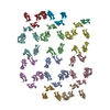
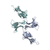
- EMDB-45459: Structure of in vitro assembled B. anthracis S-layer protein Sap -
+ Open data
- Basic information
| Entry |  | ||||||||||||
|---|---|---|---|---|---|---|---|---|---|---|---|---|---|
| Title | Structure of in vitro assembled B. anthracis S-layer protein Sap | ||||||||||||
 Map data Map data | Sharpened map from RELION postprocessing lowpass filtered to the final resolution (7.2 angstroms) | ||||||||||||
 Sample Sample |
| ||||||||||||
 Keywords Keywords | S-layer / surface protein / Bacillus anthracis / STRUCTURAL PROTEIN | ||||||||||||
| Function / homology |  Function and homology information Function and homology information | ||||||||||||
| Biological species |  | ||||||||||||
| Method | subtomogram averaging / cryo EM / Resolution: 7.2 Å | ||||||||||||
 Authors Authors | Leigh KE / Van der Verren SE / Remaut H / Kudryashev M | ||||||||||||
| Funding support |  Germany, Germany,  Belgium, 3 items Belgium, 3 items
| ||||||||||||
 Citation Citation |  Journal: Proc Natl Acad Sci U S A / Year: 2024 Journal: Proc Natl Acad Sci U S A / Year: 2024Title: Architecture of the Sap S-layer of revealed by integrative structural biology. Authors: Adrià Sogues / Kendra Leigh / Ethan V Halingstad / Sander E Van der Verren / Adam J Cecil / Antonella Fioravanti / Alexander J Pak / Misha Kudryashev / Han Remaut /    Abstract: is a spore-forming gram-positive bacterium responsible for anthrax, an infectious disease with a high mortality rate and a target of concern due to bioterrorism and long-term site contamination. The ... is a spore-forming gram-positive bacterium responsible for anthrax, an infectious disease with a high mortality rate and a target of concern due to bioterrorism and long-term site contamination. The entire surface of vegetative cells in exponential or stationary growth phase is covered in proteinaceous arrays called S-layers, composed of Sap or EA1 protein, respectively. The Sap S-layer represents an important virulence factor and cell envelope support structure whose paracrystalline nature is essential for its function. However, the spatial organization of Sap in its lattice state remains elusive. Here, we employed cryoelectron tomography and subtomogram averaging to obtain a map of the Sap S-layer from tubular polymers that revealed a conformational switch between the postassembly protomers and the previously available X-ray structure of the condensed monomers. To build and validate an atomic model of the lattice within this map, we used a combination of molecular dynamics simulations, X-ray crystallography, cross-linking mass spectrometry, and biophysics in an integrative structural biology approach. The Sap lattice model produced recapitulates a close-to-physiological arrangement, reveals high-resolution details of lattice contacts, and sheds light on the mechanisms underlying the stability of the Sap layer. | ||||||||||||
| History |
|
- Structure visualization
| Supplemental images |
|---|
- Downloads & links
-EMDB archive
| Map data |  emd_45459.map.gz emd_45459.map.gz | 24.9 MB |  EMDB map data format EMDB map data format | |
|---|---|---|---|---|
| Header (meta data) |  emd-45459-v30.xml emd-45459-v30.xml emd-45459.xml emd-45459.xml | 19.2 KB 19.2 KB | Display Display |  EMDB header EMDB header |
| FSC (resolution estimation) |  emd_45459_fsc.xml emd_45459_fsc.xml | 6.9 KB | Display |  FSC data file FSC data file |
| Images |  emd_45459.png emd_45459.png | 70.4 KB | ||
| Masks |  emd_45459_msk_1.map emd_45459_msk_1.map | 27 MB |  Mask map Mask map | |
| Filedesc metadata |  emd-45459.cif.gz emd-45459.cif.gz | 5.8 KB | ||
| Others |  emd_45459_additional_1.map.gz emd_45459_additional_1.map.gz emd_45459_half_map_1.map.gz emd_45459_half_map_1.map.gz emd_45459_half_map_2.map.gz emd_45459_half_map_2.map.gz | 25 MB 25.1 MB 25 MB | ||
| Archive directory |  http://ftp.pdbj.org/pub/emdb/structures/EMD-45459 http://ftp.pdbj.org/pub/emdb/structures/EMD-45459 ftp://ftp.pdbj.org/pub/emdb/structures/EMD-45459 ftp://ftp.pdbj.org/pub/emdb/structures/EMD-45459 | HTTPS FTP |
-Validation report
| Summary document |  emd_45459_validation.pdf.gz emd_45459_validation.pdf.gz | 1.2 MB | Display |  EMDB validaton report EMDB validaton report |
|---|---|---|---|---|
| Full document |  emd_45459_full_validation.pdf.gz emd_45459_full_validation.pdf.gz | 1.2 MB | Display | |
| Data in XML |  emd_45459_validation.xml.gz emd_45459_validation.xml.gz | 12.9 KB | Display | |
| Data in CIF |  emd_45459_validation.cif.gz emd_45459_validation.cif.gz | 17.8 KB | Display | |
| Arichive directory |  https://ftp.pdbj.org/pub/emdb/validation_reports/EMD-45459 https://ftp.pdbj.org/pub/emdb/validation_reports/EMD-45459 ftp://ftp.pdbj.org/pub/emdb/validation_reports/EMD-45459 ftp://ftp.pdbj.org/pub/emdb/validation_reports/EMD-45459 | HTTPS FTP |
-Related structure data
| Related structure data |  9g93MC  8rx2C  8s80C  8s83C M: atomic model generated by this map C: citing same article ( |
|---|---|
| Similar structure data | Similarity search - Function & homology  F&H Search F&H Search |
- Links
Links
| EMDB pages |  EMDB (EBI/PDBe) / EMDB (EBI/PDBe) /  EMDataResource EMDataResource |
|---|
- Map
Map
| File |  Download / File: emd_45459.map.gz / Format: CCP4 / Size: 27 MB / Type: IMAGE STORED AS FLOATING POINT NUMBER (4 BYTES) Download / File: emd_45459.map.gz / Format: CCP4 / Size: 27 MB / Type: IMAGE STORED AS FLOATING POINT NUMBER (4 BYTES) | ||||||||||||||||||||||||||||||||||||
|---|---|---|---|---|---|---|---|---|---|---|---|---|---|---|---|---|---|---|---|---|---|---|---|---|---|---|---|---|---|---|---|---|---|---|---|---|---|
| Annotation | Sharpened map from RELION postprocessing lowpass filtered to the final resolution (7.2 angstroms) | ||||||||||||||||||||||||||||||||||||
| Projections & slices | Image control
Images are generated by Spider. | ||||||||||||||||||||||||||||||||||||
| Voxel size | X=Y=Z: 1.8 Å | ||||||||||||||||||||||||||||||||||||
| Density |
| ||||||||||||||||||||||||||||||||||||
| Symmetry | Space group: 1 | ||||||||||||||||||||||||||||||||||||
| Details | EMDB XML:
|
-Supplemental data
-Mask #1
| File |  emd_45459_msk_1.map emd_45459_msk_1.map | ||||||||||||
|---|---|---|---|---|---|---|---|---|---|---|---|---|---|
| Projections & Slices |
| ||||||||||||
| Density Histograms |
-Additional map: Unfiltered unmasked map - average of the two half maps
| File | emd_45459_additional_1.map | ||||||||||||
|---|---|---|---|---|---|---|---|---|---|---|---|---|---|
| Annotation | Unfiltered unmasked map - average of the two half maps | ||||||||||||
| Projections & Slices |
| ||||||||||||
| Density Histograms |
-Half map: Unfiltered unmasked half map 1
| File | emd_45459_half_map_1.map | ||||||||||||
|---|---|---|---|---|---|---|---|---|---|---|---|---|---|
| Annotation | Unfiltered unmasked half map 1 | ||||||||||||
| Projections & Slices |
| ||||||||||||
| Density Histograms |
-Half map: Unfiltered unmasked half map 2
| File | emd_45459_half_map_2.map | ||||||||||||
|---|---|---|---|---|---|---|---|---|---|---|---|---|---|
| Annotation | Unfiltered unmasked half map 2 | ||||||||||||
| Projections & Slices |
| ||||||||||||
| Density Histograms |
- Sample components
-Entire : Multimeric array of B. anthracis S-layer protein Sap
| Entire | Name: Multimeric array of B. anthracis S-layer protein Sap |
|---|---|
| Components |
|
-Supramolecule #1: Multimeric array of B. anthracis S-layer protein Sap
| Supramolecule | Name: Multimeric array of B. anthracis S-layer protein Sap / type: complex / ID: 1 / Parent: 0 / Macromolecule list: all |
|---|---|
| Source (natural) | Organism:  |
-Macromolecule #1: S-layer protein Sap
| Macromolecule | Name: S-layer protein Sap / type: protein_or_peptide / ID: 1 / Enantiomer: LEVO |
|---|---|
| Source (natural) | Organism:  |
| Recombinant expression | Organism:  |
| Sequence | String: MSAKAVTTQK VEVKFSKAVE KLTKEDIKVT NKANNDKVLV KEVTLSEDKK SATVELYSNL AAKQTYTVDV NKVGKTEVAV GSLEAKTIEM ADQTVVADEP TALQFTVKDE NGTEVVSPEG IEFVTPAAEK INAKGEITLA KGTSTTVKAV YKKDGKVVAE SKEVKVSAEG ...String: MSAKAVTTQK VEVKFSKAVE KLTKEDIKVT NKANNDKVLV KEVTLSEDKK SATVELYSNL AAKQTYTVDV NKVGKTEVAV GSLEAKTIEM ADQTVVADEP TALQFTVKDE NGTEVVSPEG IEFVTPAAEK INAKGEITLA KGTSTTVKAV YKKDGKVVAE SKEVKVSAEG AAVASISNWT VAEQNKADFT SKDFKQNNKV YEGDNAYVQV ELKDQFNAVT TGKVEYESLN TEVAVVDKAT GKVTVLSAGK APVKVTVKDS KGKELVSKTV EIEAFAQKAM KEIKLEKTNV ALSTKDVTDL KVKAPVLDQY GKEFTAPVTV KVLDKDGKEL KEQKLEAKYV NKELVLNAAG QEAGNYTVVL TAKSGEKEAK ATLALELKAP GAFSKFEVRG LEKELDKYVT EENQKNAMTV SVLPVDANGL VLKGAEAAEL KVTTTNKEGK EVDATDAQVT VQNNSVITVG QGAKAGETYK VTVVLDGKLI TTHSFKVVDT APTAKGLAVE FTSTSLKEVA PNADLKAALL NILSVDGVPA TTAKATVSNV EFVSADTNVV AENGTVGAKG ATSIYVKNLT VVKDGKEQKV EFDKAVQVAV SIKEAKPATK HHHHHH UniProtKB: S-layer protein sap |
-Experimental details
-Structure determination
| Method | cryo EM |
|---|---|
 Processing Processing | subtomogram averaging |
| Aggregation state | 2D array |
- Sample preparation
| Buffer | pH: 7.4 / Details: Phosphate buffered saline |
|---|---|
| Grid | Model: UltrAuFoil R2/2 / Material: GOLD / Support film - Material: GOLD / Support film - topology: HOLEY |
| Vitrification | Cryogen name: ETHANE / Chamber humidity: 100 % / Chamber temperature: 295.15 K / Instrument: GATAN CRYOPLUNGE 3 |
- Electron microscopy
| Microscope | FEI TITAN KRIOS |
|---|---|
| Specialist optics | Energy filter - Name: GIF Quantum SE / Energy filter - Slit width: 20 eV Details: 70 micron objective aperture used in addition to the energy filter |
| Image recording | Film or detector model: GATAN K2 SUMMIT (4k x 4k) / Detector mode: SUPER-RESOLUTION / Number real images: 1 / Average electron dose: 3.8 e/Å2 |
| Electron beam | Acceleration voltage: 300 kV / Electron source:  FIELD EMISSION GUN FIELD EMISSION GUN |
| Electron optics | Illumination mode: FLOOD BEAM / Imaging mode: BRIGHT FIELD / Cs: 2.7 mm / Nominal defocus max: 3.5 µm / Nominal defocus min: 2.0 µm / Nominal magnification: 81000 |
| Sample stage | Specimen holder model: FEI TITAN KRIOS AUTOGRID HOLDER / Cooling holder cryogen: NITROGEN |
| Experimental equipment |  Model: Titan Krios / Image courtesy: FEI Company |
 Movie
Movie Controller


 Z (Sec.)
 Y (Row.)
 X (Col.)


