 Movie
Movie Controller

[English] 日本語
 Yorodumi
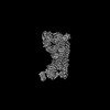
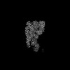
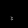
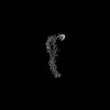
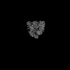
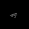
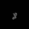
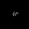
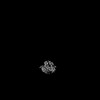
- PDB-9g08: Structure of human RNF213 bound to the secreted effector IpaH1.4 ... -
+ Open data

- Basic information
| Entry | Database: PDB / ID: 9g08 | |||||||||||||||
|---|---|---|---|---|---|---|---|---|---|---|---|---|---|---|---|---|
| Title | Structure of human RNF213 bound to the secreted effector IpaH1.4 from Shigella flexneri | |||||||||||||||
 Components Components |
| |||||||||||||||
 Keywords Keywords | ANTIMICROBIAL PROTEIN / RNF213 / IpaH1.4 / IpaH / E3 ligase / RING / LRR / NEL / secreted effector / Shigella / inhibitor / complex / AAA ATPase | |||||||||||||||
| Function / homology |  Function and homology information Function and homology informationlipid ubiquitination / negative regulation of non-canonical Wnt signaling pathway / lipid droplet formation / xenophagy / Suppression of apoptosis / sprouting angiogenesis / Transferases; Acyltransferases; Aminoacyltransferases / regulation of lipid metabolic process / protein K63-linked ubiquitination / protein autoubiquitination ...lipid ubiquitination / negative regulation of non-canonical Wnt signaling pathway / lipid droplet formation / xenophagy / Suppression of apoptosis / sprouting angiogenesis / Transferases; Acyltransferases; Aminoacyltransferases / regulation of lipid metabolic process / protein K63-linked ubiquitination / protein autoubiquitination / immune system process / lipid droplet / RING-type E3 ubiquitin transferase / Hydrolases; Acting on acid anhydrides; Acting on acid anhydrides to facilitate cellular and subcellular movement / ubiquitin-protein transferase activity / Signaling by ALK fusions and activated point mutants / ubiquitin protein ligase activity / Antigen processing: Ubiquitination & Proteasome degradation / toxin activity / angiogenesis / symbiont-mediated suppression of host NF-kappaB cascade / ubiquitin-dependent protein catabolic process / host cell cytoplasm / defense response to bacterium / protein ubiquitination / nucleolus / ATP hydrolysis activity / extracellular region / zinc ion binding / ATP binding / membrane / cytosol / cytoplasm Similarity search - Function | |||||||||||||||
| Biological species |  Homo sapiens (human) Homo sapiens (human) Shigella flexneri 5a str. M90T (bacteria) Shigella flexneri 5a str. M90T (bacteria) | |||||||||||||||
| Method | ELECTRON MICROSCOPY / single particle reconstruction / cryo EM / Resolution: 3.3 Å | |||||||||||||||
 Authors Authors | Naydenova, K. / Randow, F. | |||||||||||||||
| Funding support |  United Kingdom, 2items United Kingdom, 2items
| |||||||||||||||
 Citation Citation | Journal: Nat Struct Mol Biol / Year: 2025 Title: Shigella flexneri evades LPS ubiquitylation through IpaH1.4-mediated degradation of RNF213. Authors: Katerina Naydenova / Keith B Boyle / Claudio Pathe / Prathyush Pothukuchi / Ana Crespillo-Casado / Felix Scharte / Pierre-Mehdi Hammoudi / Elsje G Otten / Neal M Alto / Felix Randow /   Abstract: Pathogens have evolved diverse strategies to counteract host immunity. Ubiquitylation of lipopolysaccharide (LPS) on cytosol-invading bacteria by the E3 ligase RNF213 creates 'eat me' signals for ...Pathogens have evolved diverse strategies to counteract host immunity. Ubiquitylation of lipopolysaccharide (LPS) on cytosol-invading bacteria by the E3 ligase RNF213 creates 'eat me' signals for antibacterial autophagy, but whether and how cytosol-adapted bacteria avoid LPS ubiquitylation remains poorly understood. Here, we show that the enterobacterium Shigella flexneri actively antagonizes LPS ubiquitylation through IpaH1.4, a secreted effector protein with ubiquitin E3 ligase activity. IpaH1.4 binds to RNF213, ubiquitylates it and targets it for proteasomal degradation, thus counteracting host-protective LPS ubiquitylation. To understand how IpaH1.4 recognizes RNF213, we determined the cryogenic electron microscopy structure of the IpaH1.4-RNF213 complex. The specificity of the interaction is achieved through the leucine-rich repeat of IpaH1.4, which binds the RING domain of RNF213 by hijacking the conserved RING interface required for binding to ubiquitin-charged E2 enzymes. IpaH1.4 also targets other E3 ligases involved in inflammation and immunity through binding to the E2-interacting face of their RING domains, including the E3 ligase LUBAC that is required for the synthesis of M1-linked ubiquitin chains on cytosol-invading bacteria downstream of RNF213. We conclude that IpaH1.4 has evolved to antagonize multiple antibacterial and proinflammatory host E3 ligases. | |||||||||||||||
| History |
|
- Structure visualization
| Structure viewer | Molecule: MolmilJmol/JSmol |
|---|
- Downloads & links
-Download
| PDBx/mmCIF format | 9g08.cif.gz | 1.6 MB | Display | PDBx/mmCIF format |
|---|---|---|---|---|
| PDB format | pdb9g08.ent.gz | 1.3 MB | Display | PDB format |
| PDBx/mmJSON format | 9g08.json.gz | Tree view | PDBx/mmJSON format | |
| Others |  Other downloads Other downloads |
-Validation report
| Arichive directory | https://data.pdbj.org/pub/pdb/validation_reports/g0/9g08ftp://data.pdbj.org/pub/pdb/validation_reports/g0/9g08 | HTTPS FTP |
|---|
-Related structure data
| Related structure data | 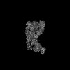 50913MC  9g09C M: map data used to model this data C: citing same article ( |
|---|---|
| Similar structure data |


-Links
 PDBj
PDBj


- Assembly
| Deposited unit | 
|
|---|---|
| 1 |
|
-Components
| #1: Protein | Mass: 596450.250 Da / Num. of mol.: 1 / Mutation: D1045N Source method: isolated from a genetically manipulated source Source: (gene. exp.) Homo sapiens (human) / Gene: RNF213, ALO17, C17orf27, KIAA1554, KIAA1618, MYSTR / Production host:   Spodoptera frugiperda (fall armyworm) Spodoptera frugiperda (fall armyworm)References: UniProt: Q63HN8, RING-type E3 ubiquitin transferase, Hydrolases; Acting on acid anhydrides; Acting on acid anhydrides to facilitate cellular and subcellular movement, Transferases; ...References: UniProt: Q63HN8, RING-type E3 ubiquitin transferase, Hydrolases; Acting on acid anhydrides; Acting on acid anhydrides to facilitate cellular and subcellular movement, Transferases; Acyltransferases; Aminoacyltransferases | ||||
|---|---|---|---|---|---|
| #2: Protein | Mass: 64925.098 Da / Num. of mol.: 1 Source method: isolated from a genetically manipulated source Source: (gene. exp.) Shigella flexneri 5a str. M90T (bacteria)Gene: ipaH1.4, SF_p0265 / Production host: References: UniProt: A0A0H2USG1, RING-type E3 ubiquitin transferase | ||||
| #3: Chemical | ChemComp-ATP /   Mass: 507.181 Da / Num. of mol.: 1 / Source method: obtained synthetically / Formula: C10H16N5O13P3 / Comment: ATP, energy-carrying molecule*YM Mass: 507.181 Da / Num. of mol.: 1 / Source method: obtained synthetically / Formula: C10H16N5O13P3 / Comment: ATP, energy-carrying molecule*YM | ||||
| #4: Chemical | ChemComp-MG /   Mass: 24.305 Da / Num. of mol.: 1 / Source method: obtained synthetically / Formula: Mg Mass: 24.305 Da / Num. of mol.: 1 / Source method: obtained synthetically / Formula: Mg | ||||
| #5: Chemical |   Mass: 65.409 Da / Num. of mol.: 3 / Source method: obtained synthetically / Formula: Zn Mass: 65.409 Da / Num. of mol.: 3 / Source method: obtained synthetically / Formula: ZnHas ligand of interest | N | Has protein modification | N | |
-Experimental details
-Experiment
| Experiment | Method: ELECTRON MICROSCOPY |
|---|---|
| EM experiment | Aggregation state: PARTICLE / 3D reconstruction method: single particle reconstruction |
- Sample preparation
| Component | Name: E3 ligase RNF213, bound to the secreted effector IpaH1.4 from Shigella flexneri Type: COMPLEX / Entity ID: #1-#2 / Source: RECOMBINANT |
|---|---|
| Molecular weight | Experimental value: NO |
| Source (natural) | Organism: Homo sapiens (human) |
| Source (recombinant) | Organism: Spodoptera frugiperda (fall armyworm) |
| Buffer solution | pH: 8 |
| Specimen | Embedding applied: NO / Shadowing applied: NO / Staining applied: NO / Vitrification applied: YES |
| Specimen support | Grid material: GOLD / Grid mesh size: 300 divisions/in. / Grid type: UltrAuFoil R1.2/1.3 |
| Vitrification | Cryogen name: ETHANE |
- Electron microscopy imaging
| Experimental equipment |  Model: Titan Krios / Image courtesy: FEI Company |
|---|---|
| Microscopy | Model: TFS KRIOS |
| Electron gun | Electron source:  FIELD EMISSION GUN / Accelerating voltage: 300 kV / Illumination mode: FLOOD BEAM FIELD EMISSION GUN / Accelerating voltage: 300 kV / Illumination mode: FLOOD BEAM |
| Electron lens | Mode: BRIGHT FIELD / Nominal defocus max: 2200 nm / Nominal defocus min: 800 nm / Calibrated defocus min: 700 nm / Calibrated defocus max: 3500 nm / Cs: 2.7 mm / C2 aperture diameter: 50 µm / Alignment procedure: COMA FREE |
| Specimen holder | Cryogen: NITROGEN / Specimen holder model: FEI TITAN KRIOS AUTOGRID HOLDER |
| Image recording | Average exposure time: 4.56 sec. / Electron dose: 32 e/Å2 / Film or detector model: FEI FALCON IV (4k x 4k) / Num. of grids imaged: 1 / Num. of real images: 16931 |
- Processing
| EM software |
| ||||||||||||||||||||||||
|---|---|---|---|---|---|---|---|---|---|---|---|---|---|---|---|---|---|---|---|---|---|---|---|---|---|
| CTF correction | Type: PHASE FLIPPING AND AMPLITUDE CORRECTION | ||||||||||||||||||||||||
| 3D reconstruction | Resolution: 3.3 Å / Resolution method: FSC 0.143 CUT-OFF / Num. of particles: 334921 / Symmetry type: POINT | ||||||||||||||||||||||||
| Refine LS restraints |
|
