 Movie
Movie Controller

[English] 日本語
 Yorodumi
- EMDB-50919: Local refinement of the RNF213 C-terminal and hinge domains in th... -
+ Open data

- Basic information
| Entry |  | |||||||||
|---|---|---|---|---|---|---|---|---|---|---|
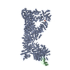
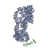
| Title | Local refinement of the RNF213 C-terminal and hinge domains in the structure of human RNF213 bound to the secreted effector IpaH1.4 from Shigella flexneri | |||||||||
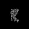
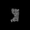
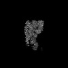
 Map data Map data | Focused refinement of the RNF213 C-terminal and hinge domains in the RNF213-IpaH1.4 complex (post-processed, auto-sharpened map) | |||||||||
 Sample Sample |
| |||||||||
 Keywords Keywords | RNF213 / IpaH1.4 / IpaH / E3 ligase / RING / LRR / NEL / secreted effector / Shigella / inhibitor / complex / AAA / ATPase / ANTIMICROBIAL PROTEIN | |||||||||
| Biological species |  Homo (humans) Homo (humans) | |||||||||
| Method | single particle reconstruction / cryo EM / Resolution: 3.1 Å | |||||||||
 Authors Authors | Naydenova K / Randow F | |||||||||
| Funding support |  United Kingdom, 2 items United Kingdom, 2 items
| |||||||||
 Citation Citation | Journal: Nat Struct Mol Biol / Year: 2025 Title: Shigella flexneri evades LPS ubiquitylation through IpaH1.4-mediated degradation of RNF213. Authors: Katerina Naydenova / Keith B Boyle / Claudio Pathe / Prathyush Pothukuchi / Ana Crespillo-Casado / Felix Scharte / Pierre-Mehdi Hammoudi / Elsje G Otten / Neal M Alto / Felix Randow /   Abstract: Pathogens have evolved diverse strategies to counteract host immunity. Ubiquitylation of lipopolysaccharide (LPS) on cytosol-invading bacteria by the E3 ligase RNF213 creates 'eat me' signals for ...Pathogens have evolved diverse strategies to counteract host immunity. Ubiquitylation of lipopolysaccharide (LPS) on cytosol-invading bacteria by the E3 ligase RNF213 creates 'eat me' signals for antibacterial autophagy, but whether and how cytosol-adapted bacteria avoid LPS ubiquitylation remains poorly understood. Here, we show that the enterobacterium Shigella flexneri actively antagonizes LPS ubiquitylation through IpaH1.4, a secreted effector protein with ubiquitin E3 ligase activity. IpaH1.4 binds to RNF213, ubiquitylates it and targets it for proteasomal degradation, thus counteracting host-protective LPS ubiquitylation. To understand how IpaH1.4 recognizes RNF213, we determined the cryogenic electron microscopy structure of the IpaH1.4-RNF213 complex. The specificity of the interaction is achieved through the leucine-rich repeat of IpaH1.4, which binds the RING domain of RNF213 by hijacking the conserved RING interface required for binding to ubiquitin-charged E2 enzymes. IpaH1.4 also targets other E3 ligases involved in inflammation and immunity through binding to the E2-interacting face of their RING domains, including the E3 ligase LUBAC that is required for the synthesis of M1-linked ubiquitin chains on cytosol-invading bacteria downstream of RNF213. We conclude that IpaH1.4 has evolved to antagonize multiple antibacterial and proinflammatory host E3 ligases. | |||||||||
| History |
|
- Structure visualization
| Supplemental images |
|---|

- Downloads & links
-EMDB archive
| Map data | emd_50919.map.gz | 12.6 MB |  EMDB map data format EMDB map data format | |
|---|---|---|---|---|
| Header (meta data) | emd-50919-v30.xmlemd-50919.xml | 18.7 KB 18.7 KB | Display Display | EMDB header |
| FSC (resolution estimation) | emd_50919_fsc.xml | 13.5 KB | Display | FSC data file |
| Images |  emd_50919.png emd_50919.png | 135.5 KB | ||
| Masks | emd_50919_msk_1.map | 216 MB | Mask map | |
| Filedesc metadata | emd-50919.cif.gz | 4.6 KB | ||
| Others | emd_50919_additional_1.map.gzemd_50919_half_map_1.map.gzemd_50919_half_map_2.map.gz | 171.4 MB 171.2 MB 171.5 MB | ||
| Archive directory |  http://ftp.pdbj.org/pub/emdb/structures/EMD-50919ftp://ftp.pdbj.org/pub/emdb/structures/EMD-50919 http://ftp.pdbj.org/pub/emdb/structures/EMD-50919ftp://ftp.pdbj.org/pub/emdb/structures/EMD-50919 | HTTPS FTP |
-Related structure data

-Links
| EMDB pages | EMDB (EBI/PDBe) / EMDataResource |
|---|
-Map
| File | Download / File: emd_50919.map.gz / Format: CCP4 / Size: 216 MB / Type: IMAGE STORED AS FLOATING POINT NUMBER (4 BYTES) | ||||||||||||||||||||||||||||||||||||
|---|---|---|---|---|---|---|---|---|---|---|---|---|---|---|---|---|---|---|---|---|---|---|---|---|---|---|---|---|---|---|---|---|---|---|---|---|---|
| Annotation | Focused refinement of the RNF213 C-terminal and hinge domains in the RNF213-IpaH1.4 complex (post-processed, auto-sharpened map) | ||||||||||||||||||||||||||||||||||||
| Projections & slices | Image control
Images are generated by Spider. | ||||||||||||||||||||||||||||||||||||
| Voxel size | X=Y=Z: 1.228 Å | ||||||||||||||||||||||||||||||||||||
| Density |
| ||||||||||||||||||||||||||||||||||||
| Symmetry | Space group: 1 | ||||||||||||||||||||||||||||||||||||
| Details | EMDB XML:
|
 Z (Sec.)
 Y (Row.)
 X (Col.)


-Supplemental data
-Mask #1
| File | emd_50919_msk_1.map | ||||||||||||
|---|---|---|---|---|---|---|---|---|---|---|---|---|---|
| Projections & Slices |
| ||||||||||||
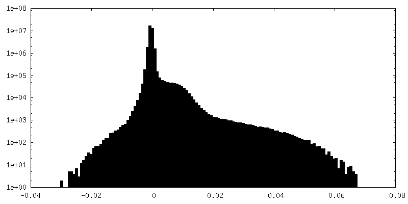
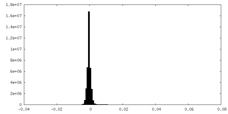
| Density Histograms |


-Additional map: Focused refinement of the RNF213 C-terminal and hinge...
| File | emd_50919_additional_1.map | ||||||||||||
|---|---|---|---|---|---|---|---|---|---|---|---|---|---|
| Annotation | Focused refinement of the RNF213 C-terminal and hinge domains in the RNF213-IpaH1.4 complex (non-filtered, non-sharpened map) | ||||||||||||
| Projections & Slices |
| ||||||||||||
| Density Histograms |


-Half map: Focused refinement of the RNF213 C-terminal and hinge...
| File | emd_50919_half_map_1.map | ||||||||||||
|---|---|---|---|---|---|---|---|---|---|---|---|---|---|
| Annotation | Focused refinement of the RNF213 C-terminal and hinge domains in the RNF213-IpaH1.4 complex (half map 1) | ||||||||||||
| Projections & Slices |
| ||||||||||||
| Density Histograms |


-Half map: Focused refinement of the RNF213 C-terminal and hinge...
| File | emd_50919_half_map_2.map | ||||||||||||
|---|---|---|---|---|---|---|---|---|---|---|---|---|---|
| Annotation | Focused refinement of the RNF213 C-terminal and hinge domains in the RNF213-IpaH1.4 complex (half map 2) | ||||||||||||
| Projections & Slices |
| ||||||||||||
| Density Histograms |


- Sample components
-Entire : E3 ligase RNF213, bound to the secreted effector IpaH1.4 from Shi...
| Entire | Name: E3 ligase RNF213, bound to the secreted effector IpaH1.4 from Shigella flexneri |
|---|---|
| Components |
|
-Supramolecule #1: E3 ligase RNF213, bound to the secreted effector IpaH1.4 from Shi...
| Supramolecule | Name: E3 ligase RNF213, bound to the secreted effector IpaH1.4 from Shigella flexneri type: complex / ID: 1 / Parent: 0 / Macromolecule list: #1-#2 |
|---|---|
| Source (natural) | Organism: Homo (humans) |
-Experimental details
-Structure determination
| Method | cryo EM |
|---|---|
 Processing Processing | single particle reconstruction |
| Aggregation state | particle |
-Sample preparation
| Buffer | pH: 8 |
|---|---|
| Grid | Model: UltrAuFoil R1.2/1.3 / Material: GOLD / Mesh: 300 / Support film - Material: GOLD / Support film - topology: HOLEY / Support film - Film thickness: 50 |
| Vitrification | Cryogen name: ETHANE |
- Electron microscopy
| Microscope | TFS KRIOS |
|---|---|
| Image recording | Film or detector model: FEI FALCON IV (4k x 4k) / Number grids imaged: 1 / Number real images: 16931 / Average exposure time: 4.56 sec. / Average electron dose: 32.0 e/Å2 |
| Electron beam | Acceleration voltage: 300 kV / Electron source:  FIELD EMISSION GUN FIELD EMISSION GUN |
| Electron optics | C2 aperture diameter: 50.0 µm / Calibrated defocus max: 3.5 µm / Calibrated defocus min: 0.7000000000000001 µm / Illumination mode: FLOOD BEAM / Imaging mode: BRIGHT FIELD / Cs: 2.7 mm / Nominal defocus max: 2.2 µm / Nominal defocus min: 0.8 µm |
| Sample stage | Specimen holder model: FEI TITAN KRIOS AUTOGRID HOLDER / Cooling holder cryogen: NITROGEN |
| Experimental equipment |  Model: Titan Krios / Image courtesy: FEI Company |

