 Movie
Movie Controller

[English] 日本語
 Yorodumi
- PDB-9fvd: Cryo-EM structure of single-layered nucleoprotein-RNA helical ass... -
+ Open data

- Basic information
| Entry | Database: PDB / ID: 9fvd | ||||||
|---|---|---|---|---|---|---|---|
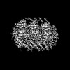
| Title | Cryo-EM structure of single-layered nucleoprotein-RNA helical assembly from Marburg virus, trimeric repeat unit | ||||||
 Components Components |
| ||||||
 Keywords Keywords | VIRAL PROTEIN / RNA-binding protein | ||||||
| Function / homology |  Function and homology information Function and homology informationviral RNA genome packaging / helical viral capsid / viral budding via host ESCRT complex / viral nucleocapsid / host cell cytoplasm / ribonucleoprotein complex / RNA binding Similarity search - Function | ||||||
| Biological species |  Marburg virus - Musoke Marburg virus - MusokeKenya 1980 | ||||||
| Method | ELECTRON MICROSCOPY / helical reconstruction / cryo EM / Resolution: 3.2 Å | ||||||
 Authors Authors | Zinzula, L. / Beck, F. / Camasta, M. / Bohn, S. / Liu, C. / Morado, D. / Bracher, A. / Plitzko, J.M. / Baumeister, W. | ||||||
| Funding support | 1items
| ||||||
 Citation Citation | Journal: Nat Commun / Year: 2024 Title: Cryo-EM structure of single-layered nucleoprotein-RNA complex from Marburg virus. Authors: Luca Zinzula / Florian Beck / Marianna Camasta / Stefan Bohn / Chuan Liu / Dustin Morado / Andreas Bracher / Juergen M Plitzko / Wolfgang Baumeister /    Abstract: Marburg virus (MARV) causes lethal hemorrhagic fever in humans, posing a threat to global health. We determined by cryogenic electron microscopy (cryo-EM) the MARV helical ribonucleoprotein (RNP) ...Marburg virus (MARV) causes lethal hemorrhagic fever in humans, posing a threat to global health. We determined by cryogenic electron microscopy (cryo-EM) the MARV helical ribonucleoprotein (RNP) complex structure in single-layered conformation, which differs from the previously reported structure of a double-layered helix. Our findings illuminate novel RNP interactions and expand knowledge on MARV genome packaging and nucleocapsid assembly, both processes representing attractive targets for the development of antiviral therapeutics against MARV disease. | ||||||
| History |
|
- Structure visualization
| Structure viewer | Molecule: MolmilJmol/JSmol |
|---|
- Downloads & links
-Download
| PDBx/mmCIF format | 9fvd.cif.gz | 248.8 KB | Display | PDBx/mmCIF format |
|---|---|---|---|---|
| PDB format | pdb9fvd.ent.gz | 205.7 KB | Display | PDB format |
| PDBx/mmJSON format | 9fvd.json.gz | Tree view | PDBx/mmJSON format | |
| Others |  Other downloads Other downloads |
-Validation report
| Arichive directory | https://data.pdbj.org/pub/pdb/validation_reports/fv/9fvdftp://data.pdbj.org/pub/pdb/validation_reports/fv/9fvd | HTTPS FTP |
|---|
-Related structure data
| Related structure data | 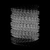 50803MC M: map data used to model this data C: citing same article ( |
|---|---|
| Similar structure data |
-Links
 PDBj
PDBj


- Assembly
| Deposited unit | 
|
|---|---|
| 1 | 
|
-Components
| #1: Protein | Mass: 49495.105 Da / Num. of mol.: 3 Source method: isolated from a genetically manipulated source Source: (gene. exp.) Marburg virus - Musoke, Kenya, 1980 / Gene: NP / Production host:  #2: RNA chain | Mass: 5816.651 Da / Num. of mol.: 5 / Source method: obtained synthetically / Source: (synth.) Marburg virus - Musoke, Kenya, 1980Has protein modification | N | |
|---|
-Experimental details
-Experiment
| Experiment | Method: ELECTRON MICROSCOPY |
|---|---|
| EM experiment | Aggregation state: HELICAL ARRAY / 3D reconstruction method: helical reconstruction |
- Sample preparation
| Component | Name: Helical assembly of nucleoprotein-RNA complex from Marburg virus Type: COMPLEX / Entity ID: all / Source: RECOMBINANT | |||||||||||||||
|---|---|---|---|---|---|---|---|---|---|---|---|---|---|---|---|---|
| Molecular weight | Experimental value: NO | |||||||||||||||
| Source (natural) | Organism: Marburg virus - Musoke, Kenya, 1980 | |||||||||||||||
| Source (recombinant) | Organism: | |||||||||||||||
| Buffer solution | pH: 8 | |||||||||||||||
| Buffer component |
| |||||||||||||||
| Specimen | Conc.: 1.25 mg/ml / Embedding applied: NO / Shadowing applied: NO / Staining applied: NO / Vitrification applied: YES | |||||||||||||||
| Specimen support | Grid material: COPPER / Grid mesh size: 400 divisions/in. / Grid type: Quantifoil R1.2/1.3 | |||||||||||||||
| Vitrification | Instrument: FEI VITROBOT MARK IV / Cryogen name: ETHANE-PROPANE / Humidity: 95 % / Chamber temperature: 277.15 K |
- Electron microscopy imaging
| Experimental equipment |  Model: Titan Krios / Image courtesy: FEI Company |
|---|---|
| Microscopy | Model: FEI TITAN KRIOS |
| Electron gun | Electron source:  FIELD EMISSION GUN / Accelerating voltage: 300 kV / Illumination mode: FLOOD BEAM FIELD EMISSION GUN / Accelerating voltage: 300 kV / Illumination mode: FLOOD BEAM |
| Electron lens | Mode: BRIGHT FIELD / Nominal defocus max: 5000 nm / Nominal defocus min: 500 nm |
| Specimen holder | Specimen holder model: FEI TITAN KRIOS AUTOGRID HOLDER |
| Image recording | Electron dose: 30 e/Å2 / Film or detector model: FEI FALCON IV (4k x 4k) / Num. of real images: 5518 |
- Processing
| EM software |
| ||||||||||||||||||||||||||||
|---|---|---|---|---|---|---|---|---|---|---|---|---|---|---|---|---|---|---|---|---|---|---|---|---|---|---|---|---|---|
| CTF correction | Type: PHASE FLIPPING AND AMPLITUDE CORRECTION | ||||||||||||||||||||||||||||
| Helical symmerty | Angular rotation/subunit: 14.732 ° / Axial rise/subunit: 2.86924 Å / Axial symmetry: C1 | ||||||||||||||||||||||||||||
| Particle selection | Num. of particles selected: 67385 | ||||||||||||||||||||||||||||
| 3D reconstruction | Resolution: 3.2 Å / Resolution method: FSC 0.143 CUT-OFF / Num. of particles: 12135 / Algorithm: FOURIER SPACE / Num. of class averages: 1 / Symmetry type: HELICAL | ||||||||||||||||||||||||||||
| Atomic model building | Protocol: AB INITIO MODEL / Space: REAL | ||||||||||||||||||||||||||||
| Atomic model building | Source name: AlphaFold / Type: in silico model |
