 Movie
Movie Controller

[English] 日本語
 Yorodumi
- PDB-9ftt: Cryo-EM structure of the type 1 pilus rod as part of the FimA-bou... -
+ Open data

- Basic information
| Entry | Database: PDB / ID: 9ftt | |||||||||||||||||||||||||||
|---|---|---|---|---|---|---|---|---|---|---|---|---|---|---|---|---|---|---|---|---|---|---|---|---|---|---|---|---|
| Title | Cryo-EM structure of the type 1 pilus rod as part of the FimA-bound usher complex (FimDHGFAnC) | |||||||||||||||||||||||||||
 Components Components | Type-1 fimbrial protein, A chain | |||||||||||||||||||||||||||
 Keywords Keywords | CELL ADHESION / chaperone / usher / pilus / rod | |||||||||||||||||||||||||||
| Function / homology |  Function and homology information Function and homology informationcell adhesion involved in single-species biofilm formation / pilus / cell adhesion / identical protein binding Similarity search - Function | |||||||||||||||||||||||||||
| Biological species |  | |||||||||||||||||||||||||||
| Method | ELECTRON MICROSCOPY / single particle reconstruction / cryo EM / Resolution: 3.6 Å | |||||||||||||||||||||||||||
 Authors Authors | Bachmann, P. / Afanasyev, P. / Boehringer, D. / Glockshuber, R. | |||||||||||||||||||||||||||
| Funding support |  Switzerland, 1items Switzerland, 1items
| |||||||||||||||||||||||||||
 Citation Citation | Journal: Nat Commun / Year: 2025 Title: Structures of the Escherichia coli type 1 pilus during pilus rod assembly and after assembly termination. Authors: Paul Bachmann / Pavel Afanasyev / Daniel Boehringer / Rudi Glockshuber / Abstract: Uropathogenic Escherichia coli strains use filamentous type 1 pili to adhere to and invade uroepithelial cells. The pilus consists of a flexible tip fibrillum, formed by the adhesin FimH and the ...Uropathogenic Escherichia coli strains use filamentous type 1 pili to adhere to and invade uroepithelial cells. The pilus consists of a flexible tip fibrillum, formed by the adhesin FimH and the subunits FimG and FimF. The pilus rod is a helical assembly of up to 3000 copies of the main subunit FimA, terminated by a single copy of the subunit FimI that anchors the rod to the assembly platform FimD in the outer membrane. Although type 1 pilus assembly can be completely reconstituted in vitro, the precise mechanism of assembly termination on FimD is still unknown. Here, we present cryo-electron microscopy structures of the fully assembled pilus with all its components prior to and after incorporation of FimI, capped with the assembly chaperone FimC. The structures reveal that FimD positions the proximal end of the pilus rod at an angle of ca. 50 degrees relative to the plane of the outer membrane. Specific interactions between FimI and FimC, absent in the equivalent FimA-FimC interface of the non-terminated pilus, stabilize the assembly-terminated state. In addition, we present structures of the transition region between the tip fibrillum and the helical rod, showing how FimF aligns the tip fibrillum along the rod axis. | |||||||||||||||||||||||||||
| History |
|
- Structure visualization
| Structure viewer | Molecule: MolmilJmol/JSmol |
|---|
- Downloads & links
-Download
| PDBx/mmCIF format | 9ftt.cif.gz | 225.2 KB | Display | PDBx/mmCIF format |
|---|---|---|---|---|
| PDB format | pdb9ftt.ent.gz | 183.9 KB | Display | PDB format |
| PDBx/mmJSON format | 9ftt.json.gz | Tree view | PDBx/mmJSON format | |
| Others |  Other downloads Other downloads |
-Validation report
| Arichive directory | https://data.pdbj.org/pub/pdb/validation_reports/ft/9fttftp://data.pdbj.org/pub/pdb/validation_reports/ft/9ftt | HTTPS FTP |
|---|
-Related structure data
| Related structure data |  50751MC  9fw9C  9fwbC  9fx0C  9fx8C  9fxaC  9fxbC 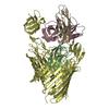 9fxsC  9fy9C M: map data used to model this data C: citing same article ( |
|---|---|
| Similar structure data |


-Links
 PDBj
PDBj
- Assembly
| Deposited unit | 
|
|---|---|
| 1 |
|
-Components
| #1: Protein | Mass: 15966.440 Da / Num. of mol.: 10 Source method: isolated from a genetically manipulated source Source: (gene. exp.) Has protein modification | Y | |
|---|
-Experimental details
-Experiment
| Experiment | Method: ELECTRON MICROSCOPY |
|---|---|
| EM experiment | Aggregation state: PARTICLE / 3D reconstruction method: single particle reconstruction |
- Sample preparation
| Component | Name: FimDHGFAnC complex / Type: COMPLEX / Entity ID: all / Source: RECOMBINANT |
|---|---|
| Molecular weight | Experimental value: NO |
| Source (natural) | Organism: |
| Source (recombinant) | Organism: |
| Buffer solution | pH: 8 |
| Specimen | Embedding applied: NO / Shadowing applied: NO / Staining applied: NO / Vitrification applied: YES |
| Specimen support | Grid material: COPPER / Grid mesh size: 300 divisions/in. / Grid type: Quantifoil R2/2 |
| Vitrification | Instrument: FEI VITROBOT MARK IV / Cryogen name: ETHANE-PROPANE / Humidity: 100 % / Chamber temperature: 277 K |
- Electron microscopy imaging
| Experimental equipment |  Model: Titan Krios / Image courtesy: FEI Company |
|---|---|
| Microscopy | Model: FEI TITAN KRIOS |
| Electron gun | Electron source:  FIELD EMISSION GUN / Accelerating voltage: 300 kV / Illumination mode: FLOOD BEAM FIELD EMISSION GUN / Accelerating voltage: 300 kV / Illumination mode: FLOOD BEAM |
| Electron lens | Mode: BRIGHT FIELD / Nominal magnification: 130000 X / Nominal defocus max: 2800 nm / Nominal defocus min: 1000 nm / Cs: 2.7 mm / C2 aperture diameter: 100 µm |
| Specimen holder | Cryogen: NITROGEN / Specimen holder model: FEI TITAN KRIOS AUTOGRID HOLDER |
| Image recording | Average exposure time: 1.1 sec. / Electron dose: 64 e/Å2 / Film or detector model: GATAN K3 BIOQUANTUM (6k x 4k) |
- Processing
| EM software |
| ||||||||||||||||||||||||
|---|---|---|---|---|---|---|---|---|---|---|---|---|---|---|---|---|---|---|---|---|---|---|---|---|---|
| CTF correction | Type: PHASE FLIPPING AND AMPLITUDE CORRECTION | ||||||||||||||||||||||||
| Symmetry | Point symmetry: C1 (asymmetric) | ||||||||||||||||||||||||
| 3D reconstruction | Resolution: 3.6 Å / Resolution method: FSC 0.143 CUT-OFF / Num. of particles: 119055 / Symmetry type: POINT | ||||||||||||||||||||||||
| Refine LS restraints |
|
