 Movie
Movie Controller

[English] 日本語
 Yorodumi
- EMDB-50846: Cryo-EM structure of the type 1 pilus rod as part of the FimI-bou... -
+ Open data

- Basic information
| Entry |  | |||||||||
|---|---|---|---|---|---|---|---|---|---|---|
| Title | Cryo-EM structure of the type 1 pilus rod as part of the FimI-bound usher complex (FimDHGFAnIC - body 1) | |||||||||
 Map data Map data | Map of the type 1 pilus rod as part of the FimI-bound usher complex FimDHGFAnIC - body 1 (sharpened) | |||||||||
 Sample Sample |
| |||||||||
 Keywords Keywords | rod / pilus / usher / termination / CELL ADHESION | |||||||||
| Function / homology |  Function and homology information Function and homology informationcell adhesion involved in single-species biofilm formation / pilus / cell adhesion / identical protein binding Similarity search - Function | |||||||||
| Biological species |  | |||||||||
| Method | single particle reconstruction / cryo EM / Resolution: 4.0 Å | |||||||||
 Authors Authors | Bachmann P / Afanasyev P / Boehringer D / Glockshuber R | |||||||||
| Funding support |  Switzerland, 1 items Switzerland, 1 items
| |||||||||
 Citation Citation | Journal: Nat Commun / Year: 2025 Title: Structures of the Escherichia coli type 1 pilus during pilus rod assembly and after assembly termination. Authors: Paul Bachmann / Pavel Afanasyev / Daniel Boehringer / Rudi Glockshuber / Abstract: Uropathogenic Escherichia coli strains use filamentous type 1 pili to adhere to and invade uroepithelial cells. The pilus consists of a flexible tip fibrillum, formed by the adhesin FimH and the ...Uropathogenic Escherichia coli strains use filamentous type 1 pili to adhere to and invade uroepithelial cells. The pilus consists of a flexible tip fibrillum, formed by the adhesin FimH and the subunits FimG and FimF. The pilus rod is a helical assembly of up to 3000 copies of the main subunit FimA, terminated by a single copy of the subunit FimI that anchors the rod to the assembly platform FimD in the outer membrane. Although type 1 pilus assembly can be completely reconstituted in vitro, the precise mechanism of assembly termination on FimD is still unknown. Here, we present cryo-electron microscopy structures of the fully assembled pilus with all its components prior to and after incorporation of FimI, capped with the assembly chaperone FimC. The structures reveal that FimD positions the proximal end of the pilus rod at an angle of ca. 50 degrees relative to the plane of the outer membrane. Specific interactions between FimI and FimC, absent in the equivalent FimA-FimC interface of the non-terminated pilus, stabilize the assembly-terminated state. In addition, we present structures of the transition region between the tip fibrillum and the helical rod, showing how FimF aligns the tip fibrillum along the rod axis. | |||||||||
| History |
|
- Structure visualization
| Supplemental images |
|---|

- Downloads & links
-EMDB archive
| Map data | emd_50846.map.gz | 96.1 MB | EMDB map data format | |
|---|---|---|---|---|
| Header (meta data) | emd-50846-v30.xmlemd-50846.xml | 22.1 KB 22.1 KB | Display Display | EMDB header |
| FSC (resolution estimation) | emd_50846_fsc.xml | 10.7 KB | Display | FSC data file |
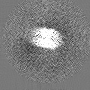
| Images |  emd_50846.png emd_50846.png | 129.8 KB | ||
| Filedesc metadata | emd-50846.cif.gz | 5.9 KB | ||
| Others | emd_50846_additional_1.map.gzemd_50846_half_map_1.map.gzemd_50846_half_map_2.map.gz | 46 MB 62.8 MB 62.8 MB | ||
| Archive directory |  http://ftp.pdbj.org/pub/emdb/structures/EMD-50846ftp://ftp.pdbj.org/pub/emdb/structures/EMD-50846 http://ftp.pdbj.org/pub/emdb/structures/EMD-50846ftp://ftp.pdbj.org/pub/emdb/structures/EMD-50846 | HTTPS FTP |
-Related structure data
| Related structure data |  9fxaMC  9fttC  9fw9C  9fwbC  9fx0C  9fx8C  9fxbC  9fxsC  9fy9C M: atomic model generated by this map C: citing same article ( |
|---|---|
| Similar structure data |


-Links
| EMDB pages | EMDB (EBI/PDBe) / EMDataResource |
|---|
-Map
| File | Download / File: emd_50846.map.gz / Format: CCP4 / Size: 103 MB / Type: IMAGE STORED AS FLOATING POINT NUMBER (4 BYTES) | ||||||||||||||||||||||||||||||||||||
|---|---|---|---|---|---|---|---|---|---|---|---|---|---|---|---|---|---|---|---|---|---|---|---|---|---|---|---|---|---|---|---|---|---|---|---|---|---|
| Annotation | Map of the type 1 pilus rod as part of the FimI-bound usher complex FimDHGFAnIC - body 1 (sharpened) | ||||||||||||||||||||||||||||||||||||
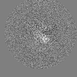
| Projections & slices | Image control
Images are generated by Spider. | ||||||||||||||||||||||||||||||||||||
| Voxel size | X=Y=Z: 1.02 Å | ||||||||||||||||||||||||||||||||||||
| Density |
| ||||||||||||||||||||||||||||||||||||
| Symmetry | Space group: 1 | ||||||||||||||||||||||||||||||||||||
| Details | EMDB XML:
|
 Z (Sec.)
 Y (Row.)
 X (Col.)


-Supplemental data
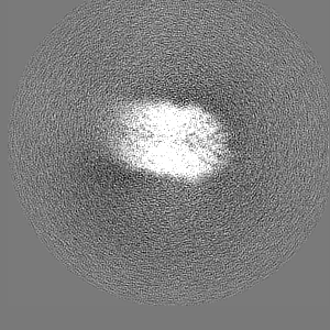
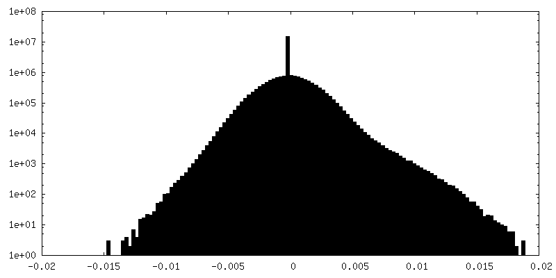
-Additional map: Map of the type 1 pilus rod as...
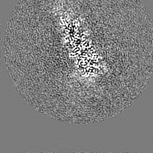
| File | emd_50846_additional_1.map | ||||||||||||
|---|---|---|---|---|---|---|---|---|---|---|---|---|---|
| Annotation | Map of the type 1 pilus rod as part of the FimI-bound usher complex FimDHGFAnIC - body 1 (unsharpened) | ||||||||||||
| Projections & Slices |
| ||||||||||||
| Density Histograms |


-Half map: Half-map A of the type 1 pilus rod...
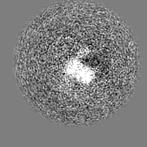
| File | emd_50846_half_map_1.map | ||||||||||||
|---|---|---|---|---|---|---|---|---|---|---|---|---|---|
| Annotation | Half-map A of the type 1 pilus rod as part of the FimI-bound usher complex FimDHGFAnIC - body 1 | ||||||||||||
| Projections & Slices |
| ||||||||||||
| Density Histograms |


-Half map: Half-map B of the type 1 pilus rod...
| File | emd_50846_half_map_2.map | ||||||||||||
|---|---|---|---|---|---|---|---|---|---|---|---|---|---|
| Annotation | Half-map B of the type 1 pilus rod as part of the FimI-bound usher complex FimDHGFAnIC - body 1 | ||||||||||||
| Projections & Slices |
| ||||||||||||
| Density Histograms |


- Sample components
-Entire : FimDHGFAnIChis complex
| Entire | Name: FimDHGFAnIChis complex |
|---|---|
| Components |
|
-Supramolecule #1: FimDHGFAnIChis complex
| Supramolecule | Name: FimDHGFAnIChis complex / type: complex / ID: 1 / Parent: 0 / Macromolecule list: all |
|---|---|
| Source (natural) | Organism: |
-Macromolecule #1: Type-1 fimbrial protein, A chain
| Macromolecule | Name: Type-1 fimbrial protein, A chain / type: protein_or_peptide / ID: 1 / Number of copies: 10 / Enantiomer: LEVO |
|---|---|
| Source (natural) | Organism: |
| Molecular weight | Theoretical: 15.96644 KDa |
| Recombinant expression | Organism: |
| Sequence | String: MAATTVNGGT VHFKGEVVNA ACAVDAGSVD QTVQLGQVRT ASLAQEGATS SAVGFNIQLN DCDTNVASKA AVAFLGTAID AGHTNVLAL QSSAAGSATN VGVQILDRTG AALTLDGATF SSETTLNNGT NTIPFQARYF ATGAATPGAA NADATFKVQY Q UniProtKB: Type-1 fimbrial protein, A chain |
-Experimental details
-Structure determination
| Method | cryo EM |
|---|---|
 Processing Processing | single particle reconstruction |
| Aggregation state | particle |
-Sample preparation
| Buffer | pH: 8 |
|---|---|
| Grid | Model: Quantifoil R2/2 / Material: COPPER / Mesh: 300 |
| Vitrification | Cryogen name: ETHANE-PROPANE / Chamber humidity: 100 % / Chamber temperature: 277 K / Instrument: FEI VITROBOT MARK IV |
- Electron microscopy
| Microscope | TFS KRIOS |
|---|---|
| Image recording | Film or detector model: GATAN K3 BIOCONTINUUM (6k x 4k) / Average exposure time: 0.6 sec. / Average electron dose: 68.0 e/Å2 |
| Electron beam | Acceleration voltage: 300 kV / Electron source:  FIELD EMISSION GUN FIELD EMISSION GUN |
| Electron optics | C2 aperture diameter: 70.0 µm / Illumination mode: FLOOD BEAM / Imaging mode: BRIGHT FIELD / Cs: 2.7 mm / Nominal defocus max: 2.4 µm / Nominal defocus min: 0.8 µm / Nominal magnification: 165000 |
| Sample stage | Specimen holder model: FEI TITAN KRIOS AUTOGRID HOLDER / Cooling holder cryogen: NITROGEN |
| Experimental equipment |  Model: Titan Krios / Image courtesy: FEI Company |

