 Movie
Movie Controller

[English] 日本語
 Yorodumi
- EMDB-50853: Cryo-EM structure of the type 1 pilus complex including pilus rod... -
+ Open data

- Basic information
| Entry |  | |||||||||
|---|---|---|---|---|---|---|---|---|---|---|
| Title | Cryo-EM structure of the type 1 pilus complex including pilus rod and FimI-bound assembly platform after incorporation of two FimI subunits - Local refinement | |||||||||
 Map data Map data | Map of the type 1 pilus complex including pilus rod and FimI-bound assembly platform after incorporation of two FimI subunits - Local refinement (sharpened) | |||||||||
 Sample Sample |
| |||||||||
 Keywords Keywords | rod / pilus / usher / termination / MEMBRANE PROTEIN | |||||||||
| Function / homology |  Function and homology information Function and homology informationfimbrial usher porin activity / pilus assembly / cell adhesion involved in single-species biofilm formation / pilus / protein folding chaperone / cell outer membrane / cell wall organization / outer membrane-bounded periplasmic space / protein folding Similarity search - Function | |||||||||
| Biological species |  | |||||||||
| Method | single particle reconstruction / cryo EM / Resolution: 4.2 Å | |||||||||
 Authors Authors | Bachmann P / Afanasyev P / Boehringer D / Glockshuber R | |||||||||
| Funding support |  Switzerland, 1 items Switzerland, 1 items
| |||||||||
 Citation Citation | Journal: Nat Commun / Year: 2025 Title: Structures of the Escherichia coli type 1 pilus during pilus rod assembly and after assembly termination. Authors: Paul Bachmann / Pavel Afanasyev / Daniel Boehringer / Rudi Glockshuber / Abstract: Uropathogenic Escherichia coli strains use filamentous type 1 pili to adhere to and invade uroepithelial cells. The pilus consists of a flexible tip fibrillum, formed by the adhesin FimH and the ...Uropathogenic Escherichia coli strains use filamentous type 1 pili to adhere to and invade uroepithelial cells. The pilus consists of a flexible tip fibrillum, formed by the adhesin FimH and the subunits FimG and FimF. The pilus rod is a helical assembly of up to 3000 copies of the main subunit FimA, terminated by a single copy of the subunit FimI that anchors the rod to the assembly platform FimD in the outer membrane. Although type 1 pilus assembly can be completely reconstituted in vitro, the precise mechanism of assembly termination on FimD is still unknown. Here, we present cryo-electron microscopy structures of the fully assembled pilus with all its components prior to and after incorporation of FimI, capped with the assembly chaperone FimC. The structures reveal that FimD positions the proximal end of the pilus rod at an angle of ca. 50 degrees relative to the plane of the outer membrane. Specific interactions between FimI and FimC, absent in the equivalent FimA-FimC interface of the non-terminated pilus, stabilize the assembly-terminated state. In addition, we present structures of the transition region between the tip fibrillum and the helical rod, showing how FimF aligns the tip fibrillum along the rod axis. | |||||||||
| History |
|
- Structure visualization
| Supplemental images |
|---|

- Downloads & links
-EMDB archive
| Map data | emd_50853.map.gz | 97.3 MB | EMDB map data format | |
|---|---|---|---|---|
| Header (meta data) | emd-50853-v30.xmlemd-50853.xml | 25.7 KB 25.7 KB | Display Display | EMDB header |
| FSC (resolution estimation) | emd_50853_fsc.xml | 11.3 KB | Display | FSC data file |
| Images |  emd_50853.png emd_50853.png | 81.1 KB | ||
| Filedesc metadata | emd-50853.cif.gz | 6.9 KB | ||
| Others | emd_50853_additional_1.map.gzemd_50853_half_map_1.map.gzemd_50853_half_map_2.map.gz | 51.6 MB 95.5 MB 95.5 MB | ||
| Archive directory |  http://ftp.pdbj.org/pub/emdb/structures/EMD-50853ftp://ftp.pdbj.org/pub/emdb/structures/EMD-50853 http://ftp.pdbj.org/pub/emdb/structures/EMD-50853ftp://ftp.pdbj.org/pub/emdb/structures/EMD-50853 | HTTPS FTP |
-Related structure data
| Related structure data | 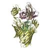 9fxsMC  9fttC  9fw9C  9fwbC  9fx0C  9fx8C  9fxaC  9fxbC  9fy9C M: atomic model generated by this map C: citing same article ( |
|---|---|
| Similar structure data |


-Links
| EMDB pages | EMDB (EBI/PDBe) / EMDataResource |
|---|
-Map
| File | Download / File: emd_50853.map.gz / Format: CCP4 / Size: 103 MB / Type: IMAGE STORED AS FLOATING POINT NUMBER (4 BYTES) | ||||||||||||||||||||||||||||||||||||
|---|---|---|---|---|---|---|---|---|---|---|---|---|---|---|---|---|---|---|---|---|---|---|---|---|---|---|---|---|---|---|---|---|---|---|---|---|---|
| Annotation | Map of the type 1 pilus complex including pilus rod and FimI-bound assembly platform after incorporation of two FimI subunits - Local refinement (sharpened) | ||||||||||||||||||||||||||||||||||||
| Projections & slices | Image control
Images are generated by Spider. | ||||||||||||||||||||||||||||||||||||
| Voxel size | X=Y=Z: 1.02 Å | ||||||||||||||||||||||||||||||||||||
| Density |
| ||||||||||||||||||||||||||||||||||||
| Symmetry | Space group: 1 | ||||||||||||||||||||||||||||||||||||
| Details | EMDB XML:
|
 Z (Sec.)
 Y (Row.)
 X (Col.)


-Supplemental data
-Additional map: Map of the type 1 pilus complex including...
| File | emd_50853_additional_1.map | ||||||||||||
|---|---|---|---|---|---|---|---|---|---|---|---|---|---|
| Annotation | Map of the type 1 pilus complex including pilus rod and FimI-bound assembly platform after incorporation of two FimI subunits - Local refinement (unsharpened) | ||||||||||||
| Projections & Slices |
| ||||||||||||
| Density Histograms |


-Half map: Half-map A of the type 1 pilus complex...
| File | emd_50853_half_map_1.map | ||||||||||||
|---|---|---|---|---|---|---|---|---|---|---|---|---|---|
| Annotation | Half-map A of the type 1 pilus complex including pilus rod and FimI-bound assembly platform after incorporation of two FimI subunits - Local refinement | ||||||||||||
| Projections & Slices |
| ||||||||||||
| Density Histograms |


-Half map: Half-map B of the type 1 pilus complex...
| File | emd_50853_half_map_2.map | ||||||||||||
|---|---|---|---|---|---|---|---|---|---|---|---|---|---|
| Annotation | Half-map B of the type 1 pilus complex including pilus rod and FimI-bound assembly platform after incorporation of two FimI subunits - Local refinement | ||||||||||||
| Projections & Slices |
| ||||||||||||
| Density Histograms |


- Sample components
-Entire : FimDHGFAnIChis complex
| Entire | Name: FimDHGFAnIChis complex |
|---|---|
| Components |
|
-Supramolecule #1: FimDHGFAnIChis complex
| Supramolecule | Name: FimDHGFAnIChis complex / type: complex / ID: 1 / Parent: 0 / Macromolecule list: all |
|---|---|
| Source (natural) | Organism: |
-Macromolecule #1: Fimbrin-like protein FimI
| Macromolecule | Name: Fimbrin-like protein FimI / type: protein_or_peptide / ID: 1 / Number of copies: 2 / Enantiomer: LEVO |
|---|---|
| Source (natural) | Organism: |
| Molecular weight | Theoretical: 17.189334 KDa |
| Recombinant expression | Organism: |
| Sequence | String: GNKWNTTLPG GNMQFQGVII AETCRIEAGD KQMTVNMGQI SSNRFHAVGE DSAPVPFVIH LRECSTVVSE RVGVAFHGVA DGKNPDVLS VGEGPGIATN IGVALFDDEG NLVPINRPPA NWKRLYSGST SLHFIAKYRA TGRRVTGGIA NAQAWFSLTY Q UniProtKB: Fimbrin-like protein FimI |
-Macromolecule #2: Chaperone protein FimC
| Macromolecule | Name: Chaperone protein FimC / type: protein_or_peptide / ID: 2 / Number of copies: 1 / Enantiomer: LEVO |
|---|---|
| Source (natural) | Organism: |
| Molecular weight | Theoretical: 23.714109 KDa |
| Recombinant expression | Organism: |
| Sequence | String: MGVALGATRV IYPAGQKQEQ LAVTNNDENS TYLIQSWVEN ADGVKDGRFI VTPPLFAMKG KKENTLRILD ATNNQLPQDR ESLFWMNVK AIPSMDKSKL TENTLQLAII SRIKLYYRPA KLALPPDQAA EKLRFRRSAN SLTLINPTPY YLTVTELNAG T RVLENALV ...String: MGVALGATRV IYPAGQKQEQ LAVTNNDENS TYLIQSWVEN ADGVKDGRFI VTPPLFAMKG KKENTLRILD ATNNQLPQDR ESLFWMNVK AIPSMDKSKL TENTLQLAII SRIKLYYRPA KLALPPDQAA EKLRFRRSAN SLTLINPTPY YLTVTELNAG T RVLENALV PPMGESTVKL PSDAGSNITY RTINDYGALT PKMTGVMEHH HHHH UniProtKB: Chaperone protein FimC |
-Macromolecule #3: Outer membrane usher protein FimD
| Macromolecule | Name: Outer membrane usher protein FimD / type: protein_or_peptide / ID: 3 / Number of copies: 1 / Enantiomer: LEVO |
|---|---|
| Source (natural) | Organism: |
| Molecular weight | Theoretical: 93.092805 KDa |
| Recombinant expression | Organism: |
| Sequence | String: DLYFNPRFLA DDPQAVADLS RFENGQELPP GTYRVDIYLN NGYMATRDVT FNTGDSEQGI VPCLTRAQLA SMGLNTASVA GMNLLADDA CVPLTTMVQD ATAHLDVGQQ RLNLTIPQAF MSNRARGYIP PELWDPGINA GLLNYNFSGN SVQNRIGGNS H YAYLNLQS ...String: DLYFNPRFLA DDPQAVADLS RFENGQELPP GTYRVDIYLN NGYMATRDVT FNTGDSEQGI VPCLTRAQLA SMGLNTASVA GMNLLADDA CVPLTTMVQD ATAHLDVGQQ RLNLTIPQAF MSNRARGYIP PELWDPGINA GLLNYNFSGN SVQNRIGGNS H YAYLNLQS GLNIGAWRLR DNTTWSYNSS DRSSGSKNKW QHINTWLERD IIPLRSRLTL GDGYTQGDIF DGINFRGAQL AS DDNMLPD SQRGFAPVIH GIARGTAQVT IKQNGYDIYN STVPPGPFTI NDIYAAGNSG DLQVTIKEAD GSTQIFTVPY SSV PLLQRE GHTRYSITAG EYRSGNAQQE KPRFFQSTLL HGLPAGWTIY GGTQLADRYR AFNFGIGKNM GALGALSVDM TQAN STLPD DSQHDGQSVR FLYNKSLNES GTNIQLVGYR YSTSGYFNFA DTTYSRMNGY NIETQDGVIQ VKPKFTDYYN LAYNK RGKL QLTVTQQLGR TSTLYLSGSH QTYWGTSNVD EQFQAGLNTA FEDINWTLSY SLTKNAWQKG RDQMLALNVN IPFSHW LRS DSKSQWRHAS ASYSMSHDLN GRMTNLAGVY GTLLEDNNLS YSVQTGYAGG GDGNSGSTGY ATLNYRGGYG NANIGYS HS DDIKQLYYGV SGGVLAHANG VTLGQPLNDT VVLVKAPGAK DAKVENQTGV RTDWRGYAVL PYATEYRENR VALDTNTL A DNVDLDNAVA NVVPTRGAIV RAEFKARVGI KLLMTLTHNN KPLPFGAMVT SESSQSSGIV ADNGQVYLSG MPLAGKVQV KWGEEENAHC VANYQLPPES QQQLLTQLSA ECRLVPRGSW SHPQFEK UniProtKB: Outer membrane usher protein FimD |
-Experimental details
-Structure determination
| Method | cryo EM |
|---|---|
 Processing Processing | single particle reconstruction |
| Aggregation state | particle |
-Sample preparation
| Buffer | pH: 8 |
|---|---|
| Grid | Model: Quantifoil R2/2 / Material: COPPER / Mesh: 300 |
| Vitrification | Cryogen name: ETHANE-PROPANE / Chamber humidity: 100 % / Chamber temperature: 277 K / Instrument: FEI VITROBOT MARK IV |
- Electron microscopy
| Microscope | TFS KRIOS |
|---|---|
| Software | Name: EPU |
| Image recording | Film or detector model: GATAN K3 BIOCONTINUUM (6k x 4k) / Average exposure time: 0.6 sec. / Average electron dose: 68.0 e/Å2 |
| Electron beam | Acceleration voltage: 300 kV / Electron source:  FIELD EMISSION GUN FIELD EMISSION GUN |
| Electron optics | C2 aperture diameter: 70.0 µm / Illumination mode: FLOOD BEAM / Imaging mode: BRIGHT FIELD / Cs: 2.7 mm / Nominal defocus max: 2.4 µm / Nominal defocus min: 0.8 µm / Nominal magnification: 165000 |
| Sample stage | Specimen holder model: FEI TITAN KRIOS AUTOGRID HOLDER / Cooling holder cryogen: NITROGEN |
| Experimental equipment |  Model: Titan Krios / Image courtesy: FEI Company |

