 Movie
Movie Controller

[English] 日本語
 Yorodumi
- PDB-9ey5: Crystal structure of human tyrosinase-related protein 1 (TYRP1) i... -
+ Open data

- Basic information
| Entry | Database: PDB / ID: 9ey5 | ||||||
|---|---|---|---|---|---|---|---|
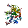
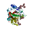
| Title | Crystal structure of human tyrosinase-related protein 1 (TYRP1) in complex with (S)-2,4-dihydroxyphenylalanine | ||||||
 Components Components | 5,6-dihydroxyindole-2-carboxylic acid oxidase | ||||||
 Keywords Keywords | METAL BINDING PROTEIN / melanogenesis / human tyrosinase / tyrosinase inhibitor | ||||||
| Function / homology |  Function and homology information Function and homology informationacetoacetic acid metabolic process / Melanin biosynthesis / Oxidoreductases; Acting on paired donors, with incorporation or reduction of molecular oxygen; With another compound as one donor, and incorporation of one atom of oxygen into the other donor / tyrosinase activity / positive regulation of melanin biosynthetic process / melanin biosynthetic process / melanocyte differentiation / melanosome organization / melanosome membrane / intracellular vesicle ...acetoacetic acid metabolic process / Melanin biosynthesis / Oxidoreductases; Acting on paired donors, with incorporation or reduction of molecular oxygen; With another compound as one donor, and incorporation of one atom of oxygen into the other donor / tyrosinase activity / positive regulation of melanin biosynthetic process / melanin biosynthetic process / melanocyte differentiation / melanosome organization / melanosome membrane / intracellular vesicle / Regulation of MITF-M-dependent genes involved in pigmentation / clathrin-coated endocytic vesicle membrane / melanosome / oxidoreductase activity / endosome membrane / protein homodimerization activity / metal ion binding / cytoplasm Similarity search - Function | ||||||
| Biological species |  Homo sapiens (human) Homo sapiens (human) | ||||||
| Method |  X-RAY DIFFRACTION / SYNCHROTRON / MOLECULAR REPLACEMENT / Resolution: 2.61 Å X-RAY DIFFRACTION / SYNCHROTRON / MOLECULAR REPLACEMENT / Resolution: 2.61 Å | ||||||
 Authors Authors | Ng, Y.M. / Soler-Lopez, M. | ||||||
| Funding support | European Union, 1items
| ||||||
 Citation Citation | Journal: Chembiochem / Year: 2024 Title: Interactions of Phenylalanine Derivatives with Human Tyrosinase: Lessons from Experimental and Theoretical tudies. Authors: Faure, C. / Min Ng, Y. / Belle, C. / Soler-Lopez, M. / Khettabi, L. / Saidi, M. / Berthet, N. / Maresca, M. / Philouze, C. / Rachidi, W. / Reglier, M. / du Moulinet d'Hardemare, A. / Jamet, H. | ||||||
| History |
|
- Structure visualization
| Structure viewer | Molecule: MolmilJmol/JSmol |
|---|
- Downloads & links
-Download
| PDBx/mmCIF format | 9ey5.cif.gz | 136.8 KB | Display | PDBx/mmCIF format |
|---|---|---|---|---|
| PDB format | pdb9ey5.ent.gz | 84.9 KB | Display | PDB format |
| PDBx/mmJSON format | 9ey5.json.gz | Tree view | PDBx/mmJSON format | |
| Others |  Other downloads Other downloads |
-Validation report
| Summary document | 9ey5_validation.pdf.gz | 2 MB | Display | wwPDB validaton report |
|---|---|---|---|---|
| Full document | 9ey5_full_validation.pdf.gz | 2 MB | Display | |
| Data in XML | 9ey5_validation.xml.gz | 20.4 KB | Display | |
| Data in CIF | 9ey5_validation.cif.gz | 27.6 KB | Display | |
| Arichive directory | https://data.pdbj.org/pub/pdb/validation_reports/ey/9ey5ftp://data.pdbj.org/pub/pdb/validation_reports/ey/9ey5 | HTTPS FTP |
-Related structure data

-Links
 PDBj
PDBj- Assembly
| Deposited unit | 
| ||||||||||||
|---|---|---|---|---|---|---|---|---|---|---|---|---|---|
| 1 |
| ||||||||||||
| Unit cell |
|
-Components
-Protein , 1 types, 1 molecules A
| #1: Protein | Mass: 50776.449 Da / Num. of mol.: 1 Source method: isolated from a genetically manipulated source Source: (gene. exp.) Homo sapiens (human) / Gene: TYRP1 / Plasmid: pACEBac1 / Cell (production host): Insect cell / Cell line (production host): Sf21 / Production host:   Spodoptera frugiperda (fall armyworm) / References: UniProt: P17643 Spodoptera frugiperda (fall armyworm) / References: UniProt: P17643 |
|---|
-Sugars , 4 types, 5 molecules 
| #2: Polysaccharide | alpha-D-mannopyranose-(1-3)-[alpha-D-mannopyranose-(1-6)]beta-D-mannopyranose-(1-4)-2-acetamido-2- ...alpha-D-mannopyranose-(1-3)-[alpha-D-mannopyranose-(1-6)]beta-D-mannopyranose-(1-4)-2-acetamido-2-deoxy-beta-D-glucopyranose-(1-4)-[alpha-L-fucopyranose-(1-6)]2-acetamido-2-deoxy-beta-D-glucopyranose Source method: isolated from a genetically manipulated source | ||
|---|---|---|---|
| #3: Polysaccharide | beta-D-mannopyranose-(1-4)-2-acetamido-2-deoxy-beta-D-glucopyranose-(1-4)-2-acetamido-2-deoxy-beta- ...beta-D-mannopyranose-(1-4)-2-acetamido-2-deoxy-beta-D-glucopyranose-(1-4)-2-acetamido-2-deoxy-beta-D-glucopyranose Source method: isolated from a genetically manipulated source | ||
| #4: Polysaccharide | Source method: isolated from a genetically manipulated source #5: Sugar | ChemComp-NAG / |  Type: D-saccharide, beta linking / Mass: 221.208 Da / Num. of mol.: 1 / Source method: obtained synthetically / Formula: C8H15NO6 Type: D-saccharide, beta linking / Mass: 221.208 Da / Num. of mol.: 1 / Source method: obtained synthetically / Formula: C8H15NO6 |
-Non-polymers , 4 types, 66 molecules 


| #6: Chemical | ChemComp-OTY /  Type: L-peptide linking / Mass: 197.188 Da / Num. of mol.: 1 / Source method: obtained synthetically / Formula: C9H11NO4 / Feature type: SUBJECT OF INVESTIGATION Type: L-peptide linking / Mass: 197.188 Da / Num. of mol.: 1 / Source method: obtained synthetically / Formula: C9H11NO4 / Feature type: SUBJECT OF INVESTIGATION | ||||
|---|---|---|---|---|---|
| #7: Chemical |  Mass: 92.094 Da / Num. of mol.: 3 / Source method: obtained synthetically / Formula: C3H8O3 Mass: 92.094 Da / Num. of mol.: 3 / Source method: obtained synthetically / Formula: C3H8O3#8: Chemical | ChemComp-ZN /  Mass: 65.409 Da / Num. of mol.: 4 / Source method: obtained synthetically / Formula: Zn Mass: 65.409 Da / Num. of mol.: 4 / Source method: obtained synthetically / Formula: Zn#9: Water | ChemComp-HOH / | Mass: 18.015 Da / Num. of mol.: 58 / Source method: isolated from a natural source / Formula: H2O |
-Details
| Has ligand of interest | Y |
|---|
-Experimental details
-Experiment
| Experiment | Method: X-RAY DIFFRACTION / Number of used crystals: 1 |
|---|
- Sample preparation
| Crystal | Density Matthews: 4.19 Å3/Da / Density % sol: 70.64 % |
|---|---|
| Crystal grow | Temperature: 277 K / Method: vapor diffusion, hanging drop / pH: 8 / Details: PEG 6000, Zinc chloride, Tris-HCl pH8.0 |
-Data collection
| Diffraction | Mean temperature: 100 K / Serial crystal experiment: N | ||||||||||||
|---|---|---|---|---|---|---|---|---|---|---|---|---|---|
| Diffraction source | Source: SYNCHROTRON / Site: ESRF  / Beamline: ID30B / Wavelength: 0.8856, 1.2782, 1.7220 / Beamline: ID30B / Wavelength: 0.8856, 1.2782, 1.7220 | ||||||||||||
| Detector | Type: DECTRIS EIGER2 X 9M / Detector: PIXEL / Date: Nov 11, 2022 | ||||||||||||
| Radiation | Monochromator: Si(111) / Protocol: MAD / Monochromatic (M) / Laue (L): M / Scattering type: x-ray | ||||||||||||
| Radiation wavelength |
| ||||||||||||
| Reflection | Resolution: 2.607→48.33 Å / Num. obs: 47641 / % possible obs: 98.63 % / Redundancy: 3.3 % / Biso Wilson estimate: 56.46 Å2 / CC1/2: 0.988 / CC star: 0.997 / Rmerge(I) obs: 0.1412 / Rpim(I) all: 0.08136 / Rrim(I) all: 0.164 / Net I/σ(I): 4.94 | ||||||||||||
| Reflection shell | Resolution: 2.61→2.7 Å / Redundancy: 3.3 % / Rmerge(I) obs: 0.6955 / Mean I/σ(I) obs: 1.01 / Num. unique obs: 4767 / CC1/2: 0.598 / CC star: 0.865 / Rpim(I) all: 0.4014 / Rrim(I) all: 0.8078 / % possible all: 98.33 |
- Processing
| Software |
| |||||||||||||||||||||||||||||||||||||||||||||||||||||||||||||||||||||||||||||
|---|---|---|---|---|---|---|---|---|---|---|---|---|---|---|---|---|---|---|---|---|---|---|---|---|---|---|---|---|---|---|---|---|---|---|---|---|---|---|---|---|---|---|---|---|---|---|---|---|---|---|---|---|---|---|---|---|---|---|---|---|---|---|---|---|---|---|---|---|---|---|---|---|---|---|---|---|---|---|
| Refinement | Method to determine structure: MOLECULAR REPLACEMENT / Resolution: 2.61→48.33 Å / SU ML: 0.328 / Cross valid method: FREE R-VALUE / σ(F): 1.33 / Phase error: 26.5752 Stereochemistry target values: GeoStd + Monomer Library + CDL v1.2
| |||||||||||||||||||||||||||||||||||||||||||||||||||||||||||||||||||||||||||||
| Solvent computation | Shrinkage radii: 0.9 Å / VDW probe radii: 1.1 Å / Solvent model: FLAT BULK SOLVENT MODEL | |||||||||||||||||||||||||||||||||||||||||||||||||||||||||||||||||||||||||||||
| Displacement parameters | Biso mean: 61.62 Å2 | |||||||||||||||||||||||||||||||||||||||||||||||||||||||||||||||||||||||||||||
| Refinement step | Cycle: LAST / Resolution: 2.61→48.33 Å
| |||||||||||||||||||||||||||||||||||||||||||||||||||||||||||||||||||||||||||||
| Refine LS restraints |
| |||||||||||||||||||||||||||||||||||||||||||||||||||||||||||||||||||||||||||||
| LS refinement shell |
|
