+ データを開く
- 基本情報
基本情報
| 登録情報 | データベース: PDB / ID: 9ewy | ||||||
|---|---|---|---|---|---|---|---|
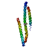
| タイトル | CryoEM structure of human MICAL1 | ||||||
 要素 要素 | [F-actin]-monooxygenase MICAL1 | ||||||
 キーワード キーワード | SIGNALING PROTEIN / MICAL / actin / actin depolymerization / axon guidance plexin / Rab | ||||||
| 機能・相同性 |  機能・相同性情報 機能・相同性情報hippocampal mossy fiber expansion / : / F-actin monooxygenase / F-actin monooxygenase activity / NAD(P)H oxidase (H2O2-forming) / sulfur oxidation / regulation of regulated secretory pathway / NAD(P)H oxidase H2O2-forming activity / oxidoreductase activity, acting on paired donors, with incorporation or reduction of molecular oxygen, NAD(P)H as one donor, and incorporation of one atom of oxygen / actin filament depolymerization ...hippocampal mossy fiber expansion / : / F-actin monooxygenase / F-actin monooxygenase activity / NAD(P)H oxidase (H2O2-forming) / sulfur oxidation / regulation of regulated secretory pathway / NAD(P)H oxidase H2O2-forming activity / oxidoreductase activity, acting on paired donors, with incorporation or reduction of molecular oxygen, NAD(P)H as one donor, and incorporation of one atom of oxygen / actin filament depolymerization / intermediate filament / actin filament bundle assembly / intercellular bridge / cytoskeleton organization / FAD binding / actin filament / monooxygenase activity / SH3 domain binding / small GTPase binding / actin filament binding / actin cytoskeleton / actin binding / Factors involved in megakaryocyte development and platelet production / midbody / endosome membrane / ciliary basal body / cilium / protein kinase binding / negative regulation of apoptotic process / signal transduction / nucleoplasm / metal ion binding / plasma membrane / cytosol / cytoplasm 類似検索 - 分子機能 | ||||||
| 生物種 |  Homo sapiens (ヒト) Homo sapiens (ヒト) | ||||||
| 手法 | 電子顕微鏡法 / 単粒子再構成法 / クライオ電子顕微鏡法 / 解像度: 3.1 Å | ||||||
 データ登録者 データ登録者 | Schrofel, A. / Pinkas, D. / Novacek, J. / Rozbesky, D. | ||||||
| 資金援助 |  チェコ, 1件 チェコ, 1件
| ||||||
 引用 引用 |  ジャーナル: Nat Commun / 年: 2024 ジャーナル: Nat Commun / 年: 2024タイトル: Structural basis of MICAL autoinhibition. 著者: Matej Horvath / Adam Schrofel / Karolina Kowalska / Jan Sabo / Jonas Vlasak / Farahdokht Nourisanami / Margarita Sobol / Daniel Pinkas / Krystof Knapp / Nicola Koupilova / Jiri Novacek / ...著者: Matej Horvath / Adam Schrofel / Karolina Kowalska / Jan Sabo / Jonas Vlasak / Farahdokht Nourisanami / Margarita Sobol / Daniel Pinkas / Krystof Knapp / Nicola Koupilova / Jiri Novacek / Vaclav Veverka / Zdenek Lansky / Daniel Rozbesky 要旨: MICAL proteins play a crucial role in cellular dynamics by binding and disassembling actin filaments, impacting processes like axon guidance, cytokinesis, and cell morphology. Their cellular activity ...MICAL proteins play a crucial role in cellular dynamics by binding and disassembling actin filaments, impacting processes like axon guidance, cytokinesis, and cell morphology. Their cellular activity is tightly controlled, as dysregulation can lead to detrimental effects on cellular morphology. Although previous studies have suggested that MICALs are autoinhibited, and require Rab proteins to become active, the detailed molecular mechanisms remained unclear. Here, we report the cryo-EM structure of human MICAL1 at a nominal resolution of 3.1 Å. Structural analyses, alongside biochemical and functional studies, show that MICAL1 autoinhibition is mediated by an intramolecular interaction between its N-terminal catalytic and C-terminal coiled-coil domains, blocking F-actin interaction. Moreover, we demonstrate that allosteric changes in the coiled-coil domain and the binding of the tripartite assembly of CH-L2α1-LIM domains to the coiled-coil domain are crucial for MICAL activation and autoinhibition. These mechanisms appear to be evolutionarily conserved, suggesting a potential universality across the MICAL family. | ||||||
| 履歴 |
|
- 構造の表示
構造の表示
| 構造ビューア | 分子:  Molmil Molmil Jmol/JSmol Jmol/JSmol |
|---|
- ダウンロードとリンク
- ダウンロード
| PDBx/mmCIF形式 |  9ewy.cif.gz 9ewy.cif.gz | 165.9 KB | 表示 |  PDBx/mmCIF形式 PDBx/mmCIF形式 |
|---|---|---|---|---|
| PDB形式 |  pdb9ewy.ent.gz pdb9ewy.ent.gz | 121.3 KB | 表示 |  PDB形式 PDB形式 |
| PDBx/mmJSON形式 |  9ewy.json.gz 9ewy.json.gz | ツリー表示 |  PDBx/mmJSON形式 PDBx/mmJSON形式 | |
| その他 |  その他のダウンロード その他のダウンロード |
-検証レポート
| 文書・要旨 |  9ewy_validation.pdf.gz 9ewy_validation.pdf.gz | 1.3 MB | 表示 |  wwPDB検証レポート wwPDB検証レポート |
|---|---|---|---|---|
| 文書・詳細版 |  9ewy_full_validation.pdf.gz 9ewy_full_validation.pdf.gz | 1.3 MB | 表示 | |
| XML形式データ |  9ewy_validation.xml.gz 9ewy_validation.xml.gz | 34.4 KB | 表示 | |
| CIF形式データ |  9ewy_validation.cif.gz 9ewy_validation.cif.gz | 49.1 KB | 表示 | |
| アーカイブディレクトリ |  https://data.pdbj.org/pub/pdb/validation_reports/ew/9ewy https://data.pdbj.org/pub/pdb/validation_reports/ew/9ewy ftp://data.pdbj.org/pub/pdb/validation_reports/ew/9ewy ftp://data.pdbj.org/pub/pdb/validation_reports/ew/9ewy | HTTPS FTP |
-関連構造データ
| 関連構造データ |  50026MC M: このデータのモデリングに利用したマップデータ C: 同じ文献を引用 ( |
|---|---|
| 類似構造データ | 類似検索 - 機能・相同性  F&H 検索 F&H 検索 |
- リンク
リンク
- 集合体
集合体
| 登録構造単位 | 
|
|---|---|
| 1 |
|
- 要素
要素
| #1: タンパク質 | [ 分子量: 118014.734 Da / 分子数: 1 / 由来タイプ: 組換発現 / 由来: (組換発現)  Homo sapiens (ヒト) / 遺伝子: MICAL1, MICAL, NICAL Homo sapiens (ヒト) / 遺伝子: MICAL1, MICAL, NICAL発現宿主:  参照: UniProt: Q8TDZ2, F-actin monooxygenase, NAD(P)H oxidase (H2O2-forming) | ||||
|---|---|---|---|---|---|
| #2: 化合物 | ChemComp-FAD / | ||||
| #3: 化合物 | | 研究の焦点であるリガンドがあるか | N | Has protein modification | N | |
-実験情報
-実験
| 実験 | 手法: 電子顕微鏡法 |
|---|---|
| EM実験 | 試料の集合状態: PARTICLE / 3次元再構成法: 単粒子再構成法 |
- 試料調製
試料調製
| 構成要素 | 名称: MICAL1 with the FAD cofactor / タイプ: COMPLEX / Entity ID: #1 / 由来: RECOMBINANT | |||||||||||||||||||||||||
|---|---|---|---|---|---|---|---|---|---|---|---|---|---|---|---|---|---|---|---|---|---|---|---|---|---|---|
| 由来(天然) | 生物種:  Homo sapiens (ヒト) Homo sapiens (ヒト) | |||||||||||||||||||||||||
| 由来(組換発現) | 生物種:  | |||||||||||||||||||||||||
| 緩衝液 | pH: 8 | |||||||||||||||||||||||||
| 緩衝液成分 |
| |||||||||||||||||||||||||
| 試料 | 濃度: 3 mg/ml / 包埋: NO / シャドウイング: NO / 染色: NO / 凍結: YES | |||||||||||||||||||||||||
| 試料支持 | グリッドの材料: GOLD / グリッドのサイズ: 300 divisions/in. / グリッドのタイプ: UltrAuFoil R1.2/1.3 | |||||||||||||||||||||||||
| 急速凍結 | 装置: FEI VITROBOT MARK IV / 凍結剤: ETHANE / 湿度: 90 % / 凍結前の試料温度: 277 K |
- 電子顕微鏡撮影
| 実験機器 |  モデル: Titan Krios / 画像提供: FEI Company |
|---|---|
| 顕微鏡 | モデル: TFS KRIOS |
| 電子銃 | 電子線源:  FIELD EMISSION GUN / 加速電圧: 300 kV / 照射モード: OTHER FIELD EMISSION GUN / 加速電圧: 300 kV / 照射モード: OTHER |
| 電子レンズ | モード: BRIGHT FIELD / 倍率(公称値): 105000 X / 倍率(補正後): 59981 X / 最大 デフォーカス(公称値): 2800 nm / 最小 デフォーカス(公称値): 1400 nm / Cs: 2.7 mm |
| 試料ホルダ | 凍結剤: NITROGEN |
| 撮影 | 平均露光時間: 2.7 sec. / 電子線照射量: 21 e/Å2 フィルム・検出器のモデル: GATAN K3 BIOQUANTUM (6k x 4k) 実像数: 59961 |
| 電子光学装置 | エネルギーフィルタースリット幅: 20 eV |
- 解析
解析
| EMソフトウェア |
| ||||||||||||||||||||||||||||||||
|---|---|---|---|---|---|---|---|---|---|---|---|---|---|---|---|---|---|---|---|---|---|---|---|---|---|---|---|---|---|---|---|---|---|
| CTF補正 | タイプ: PHASE FLIPPING AND AMPLITUDE CORRECTION | ||||||||||||||||||||||||||||||||
| 粒子像の選択 | 選択した粒子像数: 6256757 | ||||||||||||||||||||||||||||||||
| 3次元再構成 | 解像度: 3.1 Å / 解像度の算出法: FSC 0.143 CUT-OFF / 粒子像の数: 84863 / 対称性のタイプ: POINT | ||||||||||||||||||||||||||||||||
| 原子モデル構築 | B value: 77.02 / プロトコル: RIGID BODY FIT / 空間: REAL | ||||||||||||||||||||||||||||||||
| 原子モデル構築 | 3D fitting-ID: 1 / Source name: PDB / タイプ: experimental model
| ||||||||||||||||||||||||||||||||
| 精密化 | 交差検証法: NONE 立体化学のターゲット値: GeoStd + Monomer Library + CDL v1.2 | ||||||||||||||||||||||||||||||||
| 原子変位パラメータ | Biso mean: 76.71 Å2 | ||||||||||||||||||||||||||||||||
| 拘束条件 |
|
 ムービー
ムービー コントローラー


 PDBj
PDBj


