Journal: Nat Commun / Year: 2024 Title: Structural basis of MICAL autoinhibition. Authors: Matej Horvath / Adam Schrofel / Karolina Kowalska / Jan Sabo / Jonas Vlasak / Farahdokht Nourisanami / Margarita Sobol / Daniel Pinkas / Krystof Knapp / Nicola Koupilova / Jiri Novacek / ...Authors: Matej Horvath / Adam Schrofel / Karolina Kowalska / Jan Sabo / Jonas Vlasak / Farahdokht Nourisanami / Margarita Sobol / Daniel Pinkas / Krystof Knapp / Nicola Koupilova / Jiri Novacek / Vaclav Veverka / Zdenek Lansky / Daniel Rozbesky Abstract: MICAL proteins play a crucial role in cellular dynamics by binding and disassembling actin filaments, impacting processes like axon guidance, cytokinesis, and cell morphology. Their cellular activity ...MICAL proteins play a crucial role in cellular dynamics by binding and disassembling actin filaments, impacting processes like axon guidance, cytokinesis, and cell morphology. Their cellular activity is tightly controlled, as dysregulation can lead to detrimental effects on cellular morphology. Although previous studies have suggested that MICALs are autoinhibited, and require Rab proteins to become active, the detailed molecular mechanisms remained unclear. Here, we report the cryo-EM structure of human MICAL1 at a nominal resolution of 3.1 Å. Structural analyses, alongside biochemical and functional studies, show that MICAL1 autoinhibition is mediated by an intramolecular interaction between its N-terminal catalytic and C-terminal coiled-coil domains, blocking F-actin interaction. Moreover, we demonstrate that allosteric changes in the coiled-coil domain and the binding of the tripartite assembly of CH-L2α1-LIM domains to the coiled-coil domain are crucial for MICAL activation and autoinhibition. These mechanisms appear to be evolutionarily conserved, suggesting a potential universality across the MICAL family.
[F-actin]-monooxygenaseMICAL1 / Molecule interacting with CasL protein 1 / MICAL-1 / NEDD9-interacting protein with calponin ...Molecule interacting with CasL protein 1 / MICAL-1 / NEDD9-interacting protein with calponin homology and LIM domains
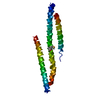
Mass: 118014.734 Da / Num. of mol.: 1 Source method: isolated from a genetically manipulated source Source: (gene. exp.) Homo sapiens (human) / Gene: MICAL1, MICAL, NICAL / Production host: Spodoptera frugiperda (fall armyworm) References: UniProt: Q8TDZ2, F-actin monooxygenase, NAD(P)H oxidase (H2O2-forming)
In the structure databanks used in Yorodumi, some data are registered as the other names, "COVID-19 virus" and "2019-nCoV". Here are the details of the virus and the list of structure data.
Jan 31, 2019. EMDB accession codes are about to change! (news from PDBe EMDB page)
EMDB accession codes are about to change! (news from PDBe EMDB page)
The allocation of 4 digits for EMDB accession codes will soon come to an end. Whilst these codes will remain in use, new EMDB accession codes will include an additional digit and will expand incrementally as the available range of codes is exhausted. The current 4-digit format prefixed with “EMD-” (i.e. EMD-XXXX) will advance to a 5-digit format (i.e. EMD-XXXXX), and so on. It is currently estimated that the 4-digit codes will be depleted around Spring 2019, at which point the 5-digit format will come into force.
The EM Navigator/Yorodumi systems omit the EMD- prefix.
Related info.:Q: What is EMD? / ID/Accession-code notation in Yorodumi/EM Navigator
Yorodumi is a browser for structure data from EMDB, PDB, SASBDB, etc.
This page is also the successor to EM Navigator detail page, and also detail information page/front-end page for Omokage search.
The word "yorodu" (or yorozu) is an old Japanese word meaning "ten thousand". "mi" (miru) is to see.
Related info.:EMDB / PDB / SASBDB / Comparison of 3 databanks / Yorodumi Search / Aug 31, 2016. New EM Navigator & Yorodumi / Yorodumi Papers / Jmol/JSmol / Function and homology information / Changes in new EM Navigator and Yorodumi
 Movie
Movie Controller


 Open data
 Basic information
 Components
 Keywords
 Function and homology information
 Homo sapiens (human)
 Authors
 Czech Republic, 1items
  Citation
 Structure visualization
 Downloads & links
 Other downloads
 PDBj
PDBj
 Assembly

 Spodoptera frugiperda (fall armyworm)
 Mass: 785.550 Da / Num. of mol.: 1 / Source method: obtained synthetically / Formula: C27H33N9O15P2 / Comment: FAD*YM
 Mass: 65.409 Da / Num. of mol.: 2 / Source method: obtained synthetically / Formula: Zn
 Sample preparation
 Electron microscopy imaging
 FIELD EMISSION GUN / Accelerating voltage: 300 kV / Illumination mode: OTHER
 Processing