 Movie
Movie Controller

[English] 日本語
 Yorodumi
- PDB-9ett: Structure of the archaellum of Sulfolobus acidocaldarius strain M... -
+ Open data

- Basic information
| Entry | Database: PDB / ID: 9ett | ||||||
|---|---|---|---|---|---|---|---|
| Title | Structure of the archaellum of Sulfolobus acidocaldarius strain MW039 (delta agl3 mutant). | ||||||
 Components Components | Flagellin | ||||||
 Keywords Keywords | PROTEIN FIBRIL / cell surface appendage / N-glycosylation / mutagenesis | ||||||
| Function / homology | archaeal-type flagellum / Flagellin, archaea / Archaebacterial flagellin / Flagellin/pilin, N-terminal / archaeal or bacterial-type flagellum-dependent cell motility / structural molecule activity / membrane / Flagellin Function and homology information Function and homology information | ||||||
| Biological species |   Sulfolobus acidocaldarius (acidophilic) Sulfolobus acidocaldarius (acidophilic) | ||||||
| Method | ELECTRON MICROSCOPY / helical reconstruction / cryo EM / Resolution: 2.37 Å | ||||||
 Authors Authors | Daum, B. / Isupov, M.N. / Gaines, M. / McLaren, M. / Mollat, C. | ||||||
| Funding support | European Union, 1items
| ||||||
 Citation Citation | Journal: Nat Commun / Year: 2024 Title: Towards a molecular picture of the archaeal cell surface. Authors: Matthew C Gaines / Michail N Isupov / Mathew McLaren / Clara L Mollat / Risat Ul Haque / Jake K Stephenson / Shamphavi Sivabalasarma / Cyril Hanus / Daniel Kattnig / Vicki A M Gold / Sonja ...Authors: Matthew C Gaines / Michail N Isupov / Mathew McLaren / Clara L Mollat / Risat Ul Haque / Jake K Stephenson / Shamphavi Sivabalasarma / Cyril Hanus / Daniel Kattnig / Vicki A M Gold / Sonja Albers / Bertram Daum /    Abstract: Archaea produce various protein filaments with specialised functions. While some archaea produce only one type of filament, the archaeal model species Sulfolobus acidocaldarius generates four. These ...Archaea produce various protein filaments with specialised functions. While some archaea produce only one type of filament, the archaeal model species Sulfolobus acidocaldarius generates four. These include rotary swimming propellers analogous to bacterial flagella (archaella), pili for twitching motility (Aap), adhesive fibres (threads), and filaments facilitating homologous recombination upon UV stress (UV pili). Here, we use cryo-electron microscopy to describe the structure of the S. acidocaldarius archaellum at 2.0 Å resolution, and update the structures of the thread and the Aap pilus at 2.7 Å and 2.6 Å resolution, respectively. We define features unique to archaella of the order Sulfolobales and compare their structure to those of Aap and threads in the context of the S-layer. We define distinct N-glycan patterns in the three filaments and identify a putative O-glycosylation site in the thread. Finally, we ascertain whether N-glycan truncation leads to structural changes in archaella and Aap. | ||||||
| History |
|
- Structure visualization
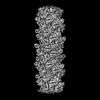
| Structure viewer | Molecule: MolmilJmol/JSmol |
|---|
- Downloads & links
-Download
| PDBx/mmCIF format | 9ett.cif.gz | 1.1 MB | Display | PDBx/mmCIF format |
|---|---|---|---|---|
| PDB format | pdb9ett.ent.gz | Display | PDB format | |
| PDBx/mmJSON format | 9ett.json.gz | Tree view | PDBx/mmJSON format | |
| Others |  Other downloads Other downloads |
-Validation report
| Arichive directory | https://data.pdbj.org/pub/pdb/validation_reports/et/9ettftp://data.pdbj.org/pub/pdb/validation_reports/et/9ett | HTTPS FTP |
|---|
-Related structure data
| Related structure data |  19961MC  8qx4C  8rzlC  9etsC  9ev0C C: citing same article ( M: map data used to model this data |
|---|---|
| Similar structure data |


-Links
 PDBj
PDBj- Assembly
| Deposited unit | 
|
|---|---|
| 1 |
|
-Components
| #1: Protein | Mass: 31209.258 Da / Num. of mol.: 20 / Source method: isolated from a natural source / Source: (natural) Sulfolobus acidocaldarius (acidophilic) / References: UniProt: Q4J9K5#2: Polysaccharide | alpha-D-mannopyranose-(1-6)-2-acetamido-2-deoxy-beta-D-glucopyranose-(1-4)-2-acetamido-2-deoxy-beta- ...alpha-D-mannopyranose-(1-6)-2-acetamido-2-deoxy-beta-D-glucopyranose-(1-4)-2-acetamido-2-deoxy-beta-D-glucopyranose Type: oligosaccharide / Mass: 586.542 Da / Num. of mol.: 120 Source method: isolated from a genetically manipulated source Has ligand of interest | Y | Has protein modification | Y | |
|---|
-Experimental details
-Experiment
| Experiment | Method: ELECTRON MICROSCOPY |
|---|---|
| EM experiment | Aggregation state: FILAMENT / 3D reconstruction method: helical reconstruction |
- Sample preparation
| Component | Name: Archaellum of the S. acidocaldarius species deficient in sulfoquinovose transferase enzyme Type: ORGANELLE OR CELLULAR COMPONENT / Entity ID: #1 / Source: NATURAL |
|---|---|
| Source (natural) | Organism: Sulfolobus acidocaldarius (acidophilic) |
| Buffer solution | pH: 3 |
| Specimen | Embedding applied: NO / Shadowing applied: NO / Staining applied: NO / Vitrification applied: YES Details: Mixed population of filaments isolated from Sulfolobus acidocaldarius deficient in agl3 - UDP-sulfoquinovose synthase |
| Vitrification | Cryogen name: ETHANE |
- Electron microscopy imaging
| Experimental equipment |  Model: Titan Krios / Image courtesy: FEI Company |
|---|---|
| Microscopy | Model: FEI TITAN KRIOS |
| Electron gun | Electron source:  FIELD EMISSION GUN / Accelerating voltage: 300 kV / Illumination mode: FLOOD BEAM FIELD EMISSION GUN / Accelerating voltage: 300 kV / Illumination mode: FLOOD BEAM |
| Electron lens | Mode: BRIGHT FIELD / Nominal defocus max: 2200 nm / Nominal defocus min: 800 nm / Cs: 2.7 mm |
| Specimen holder | Cryogen: NITROGEN / Specimen holder model: FEI TITAN KRIOS AUTOGRID HOLDER |
| Image recording | Average exposure time: 4.5 sec. / Electron dose: 40 e/Å2 / Film or detector model: TFS FALCON 4i (4k x 4k) / Num. of real images: 29189 |
- Processing
| EM software |
| ||||||||||||||||||||||||||||||||||||||||||||||||||||||||||||||||||||||||||||||||||||||||||||||||||||||||||
|---|---|---|---|---|---|---|---|---|---|---|---|---|---|---|---|---|---|---|---|---|---|---|---|---|---|---|---|---|---|---|---|---|---|---|---|---|---|---|---|---|---|---|---|---|---|---|---|---|---|---|---|---|---|---|---|---|---|---|---|---|---|---|---|---|---|---|---|---|---|---|---|---|---|---|---|---|---|---|---|---|---|---|---|---|---|---|---|---|---|---|---|---|---|---|---|---|---|---|---|---|---|---|---|---|---|---|---|
| CTF correction | Type: PHASE FLIPPING AND AMPLITUDE CORRECTION | ||||||||||||||||||||||||||||||||||||||||||||||||||||||||||||||||||||||||||||||||||||||||||||||||||||||||||
| Helical symmerty | Angular rotation/subunit: 107.9 ° / Axial rise/subunit: 5.4 Å / Axial symmetry: C1 | ||||||||||||||||||||||||||||||||||||||||||||||||||||||||||||||||||||||||||||||||||||||||||||||||||||||||||
| 3D reconstruction | Resolution: 2.37 Å / Resolution method: FSC 0.143 CUT-OFF / Num. of particles: 256869 / Symmetry type: HELICAL | ||||||||||||||||||||||||||||||||||||||||||||||||||||||||||||||||||||||||||||||||||||||||||||||||||||||||||
| Atomic model building |
| ||||||||||||||||||||||||||||||||||||||||||||||||||||||||||||||||||||||||||||||||||||||||||||||||||||||||||
| Refinement | Resolution: 2.37→2.37 Å / Cor.coef. Fo:Fc: 0.778 / SU B: 7.135 / SU ML: 0.154 / ESU R: 0.174 Stereochemistry target values: MAXIMUM LIKELIHOOD WITH PHASES
| ||||||||||||||||||||||||||||||||||||||||||||||||||||||||||||||||||||||||||||||||||||||||||||||||||||||||||
| Solvent computation | Solvent model: PARAMETERS FOR MASK CACLULATION | ||||||||||||||||||||||||||||||||||||||||||||||||||||||||||||||||||||||||||||||||||||||||||||||||||||||||||
| Displacement parameters | Biso mean: 89.706 Å2
| ||||||||||||||||||||||||||||||||||||||||||||||||||||||||||||||||||||||||||||||||||||||||||||||||||||||||||
| Refinement step | Cycle: 1 / Total: 48800 | ||||||||||||||||||||||||||||||||||||||||||||||||||||||||||||||||||||||||||||||||||||||||||||||||||||||||||
| Refine LS restraints |
|