 Movie
Movie Controller

[English] 日本語
 Yorodumi
- PDB-9ek0: Human M5 muscarinic acetylcholine receptor complex with mini-Gq a... -
+ Open data

- Basic information
| Entry | Database: PDB / ID: 9ek0 | |||||||||
|---|---|---|---|---|---|---|---|---|---|---|
| Title | Human M5 muscarinic acetylcholine receptor complex with mini-Gq and iperoxo | |||||||||
 Components Components |
| |||||||||
 Keywords Keywords | MEMBRANE PROTEIN / G protein-coupled receptor / acetylcholine binding / seven transmembrane protein | |||||||||
| Function / homology |  Function and homology information Function and homology informationregulation of phosphatidylinositol dephosphorylation / gastric acid secretion / Muscarinic acetylcholine receptors / G protein-coupled acetylcholine receptor activity / phosphatidylinositol-4,5-bisphosphate phospholipase C activity / adenylate cyclase-inhibiting G protein-coupled acetylcholine receptor signaling pathway / G-protein activation / Activation of the phototransduction cascade / Glucagon-type ligand receptors / Thromboxane signalling through TP receptor ...regulation of phosphatidylinositol dephosphorylation / gastric acid secretion / Muscarinic acetylcholine receptors / G protein-coupled acetylcholine receptor activity / phosphatidylinositol-4,5-bisphosphate phospholipase C activity / adenylate cyclase-inhibiting G protein-coupled acetylcholine receptor signaling pathway / G-protein activation / Activation of the phototransduction cascade / Glucagon-type ligand receptors / Thromboxane signalling through TP receptor / Sensory perception of sweet, bitter, and umami (glutamate) taste / G beta:gamma signalling through PI3Kgamma / G beta:gamma signalling through CDC42 / Cooperation of PDCL (PhLP1) and TRiC/CCT in G-protein beta folding / Activation of G protein gated Potassium channels / Inhibition of voltage gated Ca2+ channels via Gbeta/gamma subunits / Ca2+ pathway / G alpha (z) signalling events / High laminar flow shear stress activates signaling by PIEZO1 and PECAM1:CDH5:KDR in endothelial cells / Glucagon-like Peptide-1 (GLP1) regulates insulin secretion / Vasopressin regulates renal water homeostasis via Aquaporins / Adrenaline,noradrenaline inhibits insulin secretion / ADP signalling through P2Y purinoceptor 12 / G alpha (q) signalling events / G alpha (i) signalling events / Thrombin signalling through proteinase activated receptors (PARs) / photoreceptor outer segment membrane / spectrin binding / alkylglycerophosphoethanolamine phosphodiesterase activity / sensory perception of taste / photoreceptor outer segment / G protein-coupled receptor signaling pathway, coupled to cyclic nucleotide second messenger / retina development in camera-type eye / cardiac muscle cell apoptotic process / photoreceptor inner segment / cell population proliferation / phospholipase C-activating G protein-coupled receptor signaling pathway / G protein-coupled acetylcholine receptor signaling pathway / G beta:gamma signalling through PLC beta / Presynaptic function of Kainate receptors / Thromboxane signalling through TP receptor / Activation of G protein gated Potassium channels / Inhibition of voltage gated Ca2+ channels via Gbeta/gamma subunits / G-protein activation / Glucagon signaling in metabolic regulation / G beta:gamma signalling through CDC42 / Prostacyclin signalling through prostacyclin receptor / G beta:gamma signalling through BTK / ADP signalling through P2Y purinoceptor 12 / Glucagon-type ligand receptors / Adrenaline,noradrenaline inhibits insulin secretion / Vasopressin regulates renal water homeostasis via Aquaporins / Glucagon-like Peptide-1 (GLP1) regulates insulin secretion / G alpha (z) signalling events / cellular response to catecholamine stimulus / ADP signalling through P2Y purinoceptor 1 / G beta:gamma signalling through PI3Kgamma / ADORA2B mediated anti-inflammatory cytokines production / adenylate cyclase-activating dopamine receptor signaling pathway / Cooperation of PDCL (PhLP1) and TRiC/CCT in G-protein beta folding / cellular response to prostaglandin E stimulus / GPER1 signaling / heterotrimeric G-protein complex / G alpha (12/13) signalling events / G-protein beta-subunit binding / positive regulation of cytosolic calcium ion concentration / Thrombin signalling through proteinase activated receptors (PARs) / signaling receptor complex adaptor activity / cell body / GTPase binding / chemical synaptic transmission / cellular response to hypoxia / Ca2+ pathway / High laminar flow shear stress activates signaling by PIEZO1 and PECAM1:CDH5:KDR in endothelial cells / G alpha (i) signalling events / G alpha (s) signalling events / G alpha (q) signalling events / postsynaptic membrane / Extra-nuclear estrogen signaling / G protein-coupled receptor signaling pathway / GTPase activity / synapse / dendrite / protein-containing complex binding / extracellular exosome / membrane / plasma membrane / cytoplasm Similarity search - Function | |||||||||
| Biological species |  Homo sapiens (human) Homo sapiens (human) | |||||||||
| Method | ELECTRON MICROSCOPY / single particle reconstruction / cryo EM / Resolution: 2.79 Å | |||||||||
 Authors Authors | Burger, W.A.C. / Mobbs, J.I. / Thal, D.M. | |||||||||
| Funding support |  Australia, 2items Australia, 2items
| |||||||||
 Citation Citation | Journal: Nat Commun / Year: 2025 Title: Cryo-EM reveals an extrahelical allosteric binding site at the M mAChR. Authors: Wessel A C Burger / Jesse I Mobbs / Bhavika Rana / Jinan Wang / Keya Joshi / Patrick R Gentry / Mahmuda Yeasmin / Hariprasad Venugopal / Aaron M Bender / Craig W Lindsley / Yinglong Miao / ...Authors: Wessel A C Burger / Jesse I Mobbs / Bhavika Rana / Jinan Wang / Keya Joshi / Patrick R Gentry / Mahmuda Yeasmin / Hariprasad Venugopal / Aaron M Bender / Craig W Lindsley / Yinglong Miao / Arthur Christopoulos / Celine Valant / David M Thal /  Abstract: The M muscarinic acetylcholine receptor (M mAChR) represents a promising therapeutic target for neurological disorders. However, the high conservation of its orthosteric binding site poses ...The M muscarinic acetylcholine receptor (M mAChR) represents a promising therapeutic target for neurological disorders. However, the high conservation of its orthosteric binding site poses significant challenges for drug development. While selective positive allosteric modulators (PAMs) offer a potential solution, a structural understanding of the M mAChR and its allosteric binding sites remains limited. Here, we present a 2.8 Å cryo-electron microscopy structure of the M mAChR complexed with heterotrimeric G protein and the agonist iperoxo, completing the active-state structural characterization of the mAChR family. To identify the binding site of M-selective PAMs, we implement an integrated approach combining mutagenesis, pharmacological assays, structural biology, and molecular dynamics simulations. Our mutagenesis studies reveal that selective M PAMs bind outside previously characterized M mAChR allosteric sites. Subsequently, we obtain a 2.1 Å structure of M mAChR co-bound with acetylcholine and the selective PAM VU6007678, revealing an allosteric pocket at the extrahelical interface between transmembrane domains 3 and 4 that is confirmed through mutagenesis and simulations. These findings demonstrate the diverse mechanisms of allosteric regulation in mAChRs and highlight the value of integrating pharmacological and structural approaches to identify allosteric binding sites. | |||||||||
| History |
|
- Structure visualization
| Structure viewer | Molecule: MolmilJmol/JSmol |
|---|
- Downloads & links
-Download
| PDBx/mmCIF format | 9ek0.cif.gz | 191.2 KB | Display | PDBx/mmCIF format |
|---|---|---|---|---|
| PDB format | pdb9ek0.ent.gz | 145.5 KB | Display | PDB format |
| PDBx/mmJSON format | 9ek0.json.gz | Tree view | PDBx/mmJSON format | |
| Others |  Other downloads Other downloads |
-Validation report
| Arichive directory | https://data.pdbj.org/pub/pdb/validation_reports/ek/9ek0ftp://data.pdbj.org/pub/pdb/validation_reports/ek/9ek0 | HTTPS FTP |
|---|
-Related structure data
| Related structure data |  48111MC  9ejzC M: map data used to model this data C: citing same article ( |
|---|---|
| Similar structure data |


-Links
 PDBj
PDBj


- Assembly
| Deposited unit | 
|
|---|---|
| 1 |
|
-Components
| #1: Protein | Mass: 39190.051 Da / Num. of mol.: 1 Source method: isolated from a genetically manipulated source Source: (gene. exp.) Homo sapiens (human) / Gene: CHRM5 / Production host:   Spodoptera frugiperda (fall armyworm) / References: UniProt: P08912 Spodoptera frugiperda (fall armyworm) / References: UniProt: P08912 |
|---|---|
| #2: Protein | Mass: 38534.062 Da / Num. of mol.: 1 Source method: isolated from a genetically manipulated source Source: (gene. exp.) Trichoplusia ni (cabbage looper) / References: UniProt: P54311 |
| #3: Protein | Mass: 28039.793 Da / Num. of mol.: 1 Source method: isolated from a genetically manipulated source Source: (gene. exp.) Homo sapiens (human) / Production host: Trichoplusia ni (cabbage looper) |
| #4: Protein | Mass: 7563.750 Da / Num. of mol.: 1 Source method: isolated from a genetically manipulated source Source: (gene. exp.) Homo sapiens (human) / Gene: GNG2 / Production host: Trichoplusia ni (cabbage looper) / References: UniProt: P59768 |
| #5: Chemical | ChemComp-IXO / 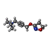  Mass: 197.254 Da / Num. of mol.: 1 / Source method: obtained synthetically / Formula: C10H17N2O2 / Feature type: SUBJECT OF INVESTIGATION Mass: 197.254 Da / Num. of mol.: 1 / Source method: obtained synthetically / Formula: C10H17N2O2 / Feature type: SUBJECT OF INVESTIGATION |
| Has ligand of interest | Y |
| Has protein modification | Y |
-Experimental details
-Experiment
| Experiment | Method: ELECTRON MICROSCOPY |
|---|---|
| EM experiment | Aggregation state: PARTICLE / 3D reconstruction method: single particle reconstruction |
- Sample preparation
| Component | Name: Human M5-miniGq-Gb1g2 complex / Type: COMPLEX / Entity ID: #1-#4 / Source: RECOMBINANT |
|---|---|
| Molecular weight | Experimental value: NO |
| Source (natural) | Organism: Homo sapiens (human) |
| Source (recombinant) | Organism: Spodoptera frugiperda (fall armyworm) |
| Buffer solution | pH: 7.5 |
| Specimen | Embedding applied: NO / Shadowing applied: NO / Staining applied: NO / Vitrification applied: YES |
| Vitrification | Cryogen name: ETHANE |
- Electron microscopy imaging
| Experimental equipment |  Model: Titan Krios / Image courtesy: FEI Company |
|---|---|
| Microscopy | Model: TFS KRIOS |
| Electron gun | Electron source:  FIELD EMISSION GUN / Accelerating voltage: 300 kV / Illumination mode: FLOOD BEAM FIELD EMISSION GUN / Accelerating voltage: 300 kV / Illumination mode: FLOOD BEAM |
| Electron lens | Mode: BRIGHT FIELD / Nominal defocus max: 1500 nm / Nominal defocus min: 500 nm |
| Image recording | Electron dose: 60 e/Å2 / Film or detector model: GATAN K3 (6k x 4k) |
- Processing
| EM software | Name: PHENIX / Version: 1.21rc1_5127 / Category: model refinement | ||||||||||||||||||||||||
|---|---|---|---|---|---|---|---|---|---|---|---|---|---|---|---|---|---|---|---|---|---|---|---|---|---|
| CTF correction | Type: PHASE FLIPPING AND AMPLITUDE CORRECTION | ||||||||||||||||||||||||
| 3D reconstruction | Resolution: 2.79 Å / Resolution method: FSC 0.143 CUT-OFF / Num. of particles: 426714 / Symmetry type: POINT | ||||||||||||||||||||||||
| Refinement | Highest resolution: 2.79 Å Stereochemistry target values: REAL-SPACE (WEIGHTED MAP SUM AT ATOM CENTERS) | ||||||||||||||||||||||||
| Refine LS restraints |
|
