 Movie
Movie Controller

[English] 日本語
 Yorodumi
- PDB-9ebw: Chimeric fluorescence biosensor formed from a lactate-binding pro... -
+ Open data

- Basic information
| Entry | Database: PDB / ID: 9ebw | ||||||||||||
|---|---|---|---|---|---|---|---|---|---|---|---|---|---|
| Title | Chimeric fluorescence biosensor formed from a lactate-binding protein and GFP, bound to lactate | ||||||||||||
 Components Components | Green fluorescent protein,Methyl-accepting chemotaxis transducer (TlpC) | ||||||||||||
 Keywords Keywords | FLUORESCENT PROTEIN / Biosensor / Binding protein / Chimera | ||||||||||||
| Function / homology |  Function and homology information Function and homology informationbioluminescence / generation of precursor metabolites and energy / chemotaxis / signal transduction / membrane Similarity search - Function | ||||||||||||
| Biological species |   Aequorea victoria (jellyfish) Aequorea victoria (jellyfish)  Helicobacter pylori (bacteria) Helicobacter pylori (bacteria) | ||||||||||||
| Method |  X-RAY DIFFRACTION / SYNCHROTRON / MOLECULAR REPLACEMENT / Resolution: 2.78 Å X-RAY DIFFRACTION / SYNCHROTRON / MOLECULAR REPLACEMENT / Resolution: 2.78 Å | ||||||||||||
 Authors Authors | Horwitz, S.M. / Ambarian, J.A. / Jones, R. / Waidmann, L. / Davis, K.M. | ||||||||||||
| Funding support |  United States, 3items United States, 3items
| ||||||||||||
 Citation Citation | Journal: Proc.Natl.Acad.Sci.USA / Year: 2025 Title: State-dependent motion of a genetically encoded fluorescent biosensor. Authors: Rosen, P.C. / Horwitz, S.M. / Brooks, D.J. / Kim, E. / Ambarian, J.A. / Waidmann, L. / Davis, K.M. / Yellen, G. | ||||||||||||
| History |
|
- Structure visualization
| Structure viewer | Molecule: MolmilJmol/JSmol |
|---|
- Downloads & links
-Download
| PDBx/mmCIF format | 9ebw.cif.gz | 376.9 KB | Display | PDBx/mmCIF format |
|---|---|---|---|---|
| PDB format | pdb9ebw.ent.gz | 301.2 KB | Display | PDB format |
| PDBx/mmJSON format | 9ebw.json.gz | Tree view | PDBx/mmJSON format | |
| Others |  Other downloads Other downloads |
-Validation report
| Arichive directory | https://data.pdbj.org/pub/pdb/validation_reports/eb/9ebwftp://data.pdbj.org/pub/pdb/validation_reports/eb/9ebw | HTTPS FTP |
|---|
-Related structure data

-Links
 PDBj
PDBj


- Assembly
| Deposited unit | 
| ||||||||
|---|---|---|---|---|---|---|---|---|---|
| 1 | 
| ||||||||
| 2 | 
| ||||||||
| Unit cell |
|
-Components
| #1: Protein | Mass: 58440.969 Da / Num. of mol.: 4 Source method: isolated from a genetically manipulated source Details: mTurquoise GFP (residues 10-153 and 428-515) attached to lactate binding protein TlpC (residues 159-421) by two separate linkers, one from residues 154-158 and another from residues 425-427 Source: (gene. exp.) Aequorea victoria (jellyfish), (gene. exp.) Helicobacter pylori (bacteria)Gene: GFP, HP_0082 / Strain: 26695 / Production host: #2: Chemical |   Mass: 92.094 Da / Num. of mol.: 2 / Source method: obtained synthetically / Formula: C3H8O3 Mass: 92.094 Da / Num. of mol.: 2 / Source method: obtained synthetically / Formula: C3H8O3#3: Chemical | 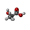  Mass: 90.078 Da / Num. of mol.: 2 / Source method: obtained synthetically / Formula: C3H6O3 / Feature type: SUBJECT OF INVESTIGATION Mass: 90.078 Da / Num. of mol.: 2 / Source method: obtained synthetically / Formula: C3H6O3 / Feature type: SUBJECT OF INVESTIGATION#4: Water | ChemComp-HOH / |  Mass: 18.015 Da / Num. of mol.: 119 / Source method: isolated from a natural source / Formula: H2O Mass: 18.015 Da / Num. of mol.: 119 / Source method: isolated from a natural source / Formula: H2OHas ligand of interest | Y | Has protein modification | Y | |
|---|
-Experimental details
-Experiment
| Experiment | Method: X-RAY DIFFRACTION / Number of used crystals: 1 |
|---|
- Sample preparation
| Crystal | Density Matthews: 2.5 Å3/Da / Density % sol: 47.4 % / Description: Green needles, 100 x 25 uM |
|---|---|
| Crystal grow | Temperature: 296 K / Method: vapor diffusion, sitting drop / Details: citric acid, PEG 6000, 1,3-propanediol |
-Data collection
| Diffraction | Mean temperature: 100 K / Serial crystal experiment: N |
|---|---|
| Diffraction source | Source: SYNCHROTRON / Site: NSLS-II / Beamline: 17-ID-1 / Wavelength: 0.920119 Å |
| Detector | Type: DECTRIS EIGER X 9M / Detector: PIXEL / Date: Mar 3, 2024 |
| Radiation | Protocol: SINGLE WAVELENGTH / Monochromatic (M) / Laue (L): M / Scattering type: x-ray |
| Radiation wavelength | Wavelength: 0.920119 Å / Relative weight: 1 |
| Reflection | Resolution: 2.78→45.94 Å / Num. obs: 55034 / % possible obs: 95.44 % / Redundancy: 4.6 % / CC1/2: 0.992 / CC star: 0.998 / Rmerge(I) obs: 0.1481 / Rpim(I) all: 0.07007 / Net I/σ(I): 7.4 |
| Reflection shell | Resolution: 2.78→2.88 Å / Rmerge(I) obs: 0.6888 / Mean I/σ(I) obs: 1.82 / Num. unique obs: 5330 / CC1/2: 0.826 / CC star: 0.951 / Rpim(I) all: 0.3447 / Rrim(I) all: 0.7748 |
- Processing
| Software |
| |||||||||||||||||||||||||||||||||||||||||||||||||||||||||||||||||||||||||||||||||||||||||||||||||||||||||
|---|---|---|---|---|---|---|---|---|---|---|---|---|---|---|---|---|---|---|---|---|---|---|---|---|---|---|---|---|---|---|---|---|---|---|---|---|---|---|---|---|---|---|---|---|---|---|---|---|---|---|---|---|---|---|---|---|---|---|---|---|---|---|---|---|---|---|---|---|---|---|---|---|---|---|---|---|---|---|---|---|---|---|---|---|---|---|---|---|---|---|---|---|---|---|---|---|---|---|---|---|---|---|---|---|---|---|
| Refinement | Method to determine structure: MOLECULAR REPLACEMENT / Resolution: 2.78→45.94 Å / SU ML: 0.45 / Cross valid method: FREE R-VALUE / σ(F): 1.33 / Phase error: 34.05 / Stereochemistry target values: ML
| |||||||||||||||||||||||||||||||||||||||||||||||||||||||||||||||||||||||||||||||||||||||||||||||||||||||||
| Solvent computation | Shrinkage radii: 0.9 Å / VDW probe radii: 1.1 Å / Solvent model: FLAT BULK SOLVENT MODEL | |||||||||||||||||||||||||||||||||||||||||||||||||||||||||||||||||||||||||||||||||||||||||||||||||||||||||
| Refinement step | Cycle: LAST / Resolution: 2.78→45.94 Å
| |||||||||||||||||||||||||||||||||||||||||||||||||||||||||||||||||||||||||||||||||||||||||||||||||||||||||
| Refine LS restraints |
| |||||||||||||||||||||||||||||||||||||||||||||||||||||||||||||||||||||||||||||||||||||||||||||||||||||||||
| LS refinement shell |
|
