Movie
Movie Controller


+ Open data

- Basic information
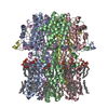
| Entry | Database: PDB / ID: 9dyk | ||||||||||||||||||||||||
|---|---|---|---|---|---|---|---|---|---|---|---|---|---|---|---|---|---|---|---|---|---|---|---|---|---|
| Title | BEST2 + GABA closed state | ||||||||||||||||||||||||
 Components Components | Bestrophin-2 | ||||||||||||||||||||||||
 Keywords Keywords | MEMBRANE PROTEIN / calcium-activated chloride channel / GABA-bound anion channel / channel-activator complex | ||||||||||||||||||||||||
| Function / homology |  Function and homology information Function and homology informationintracellularly ligand-gated monoatomic ion channel activity / ligand-gated monoatomic anion channel activity / bicarbonate channel activity / ligand-gated monoatomic cation channel activity / chloride channel activity / membrane depolarization / chloride channel complex / chloride transmembrane transport / Stimuli-sensing channels / sensory perception of smell ...intracellularly ligand-gated monoatomic ion channel activity / ligand-gated monoatomic anion channel activity / bicarbonate channel activity / ligand-gated monoatomic cation channel activity / chloride channel activity / membrane depolarization / chloride channel complex / chloride transmembrane transport / Stimuli-sensing channels / sensory perception of smell / basolateral plasma membrane / cilium / metal ion binding / plasma membrane Similarity search - Function | ||||||||||||||||||||||||
| Biological species |  Homo sapiens (human) Homo sapiens (human) | ||||||||||||||||||||||||
| Method | ELECTRON MICROSCOPY / single particle reconstruction / cryo EM / Resolution: 2.27 Å | ||||||||||||||||||||||||
 Authors Authors | Owji, A.P. / Kittredge, A. / Zhang, Y. / Yang, T. | ||||||||||||||||||||||||
| Funding support |  United States, 7items United States, 7items
| ||||||||||||||||||||||||
 Citation Citation | Journal: Nat Commun / Year: 2024 Title: Neurotransmitter-bound bestrophin channel structures reveal small molecule drug targeting sites for disease treatment. Authors: Aaron P Owji / Jingyun Dong / Alec Kittredge / Jiali Wang / Yu Zhang / Tingting Yang / Abstract: Best1 and Best2 are two members of the bestrophin family of anion channels critically involved in the prevention of retinal degeneration and maintenance of intraocular pressure, respectively. Here, ...Best1 and Best2 are two members of the bestrophin family of anion channels critically involved in the prevention of retinal degeneration and maintenance of intraocular pressure, respectively. Here, we solved glutamate- and γ-aminobutyric acid (GABA)-bound Best2 structures, which delineate an intracellular glutamate binding site and an extracellular GABA binding site on Best2, respectively, identified extracellular GABA as a permeable activator of Best2, and elucidated the co-regulation of Best2 by glutamate, GABA and glutamine synthetase in vivo. We further identified multiple small molecules as activators of the bestrophin channels. Extensive analyses were carried out for a potent activator, 4-aminobenzoic acid (PABA): PABA-bound Best1 and Best2 structures are solved and illustrate the same binding site as in GABA-bound Best2; PABA treatment rescues the functional deficiency of patient-derived Best1 mutations. Together, our results demonstrate the mechanism and potential of multiple small molecule candidates as clinically applicable drugs for bestrophin-associated diseases/conditions. | ||||||||||||||||||||||||
| History |
|
- Structure visualization
| Structure viewer | Molecule: MolmilJmol/JSmol |
|---|
- Downloads & links
-Download
| PDBx/mmCIF format | 9dyk.cif.gz | 332.3 KB | Display | PDBx/mmCIF format |
|---|---|---|---|---|
| PDB format | pdb9dyk.ent.gz | 276.3 KB | Display | PDB format |
| PDBx/mmJSON format | 9dyk.json.gz | Tree view | PDBx/mmJSON format | |
| Others |  Other downloads Other downloads |
-Validation report
| Arichive directory | https://data.pdbj.org/pub/pdb/validation_reports/dy/9dykftp://data.pdbj.org/pub/pdb/validation_reports/dy/9dyk | HTTPS FTP |
|---|
-Related structure data
| Related structure data |  47307MC 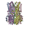 9dyhC  9dyiC 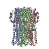 9dyjC 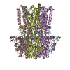 9dylC 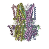 9dymC 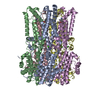 9dynC 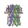 9dyoC M: map data used to model this data C: citing same article ( |
|---|---|
| Similar structure data |
-Links
 PDBj
PDBj- Assembly
| Deposited unit | 
|
|---|---|
| 1 |
|
-Components
| #1: Protein | Mass: 46796.898 Da / Num. of mol.: 5 Source method: isolated from a genetically manipulated source Source: (gene. exp.) Homo sapiens (human) / Gene: BEST2, VMD2L1 / Cell line (production host): HEK293 / Production host: Homo sapiens (human) / References: UniProt: Q8NFU1#2: Chemical | ChemComp-CA /   Mass: 40.078 Da / Num. of mol.: 5 / Source method: obtained synthetically / Formula: Ca Mass: 40.078 Da / Num. of mol.: 5 / Source method: obtained synthetically / Formula: Ca#3: Chemical | ChemComp-CL / |   Mass: 35.453 Da / Num. of mol.: 1 / Source method: obtained synthetically / Formula: Cl Mass: 35.453 Da / Num. of mol.: 1 / Source method: obtained synthetically / Formula: ClHas ligand of interest | N | Has protein modification | N | |
|---|
-Experimental details
-Experiment
| Experiment | Method: ELECTRON MICROSCOPY |
|---|---|
| EM experiment | Aggregation state: PARTICLE / 3D reconstruction method: single particle reconstruction |
- Sample preparation
| Component | Name: BEST2 + GABA closed state / Type: COMPLEX / Entity ID: #1 / Source: RECOMBINANT | ||||||||||||||||||||
|---|---|---|---|---|---|---|---|---|---|---|---|---|---|---|---|---|---|---|---|---|---|
| Molecular weight | Value: 0.286 MDa / Experimental value: NO | ||||||||||||||||||||
| Source (natural) | Organism: Homo sapiens (human) | ||||||||||||||||||||
| Source (recombinant) | Organism: Homo sapiens (human) / Cell: HEK293 | ||||||||||||||||||||
| Buffer solution | pH: 7.8 | ||||||||||||||||||||
| Buffer component |
| ||||||||||||||||||||
| Specimen | Conc.: 5 mg/ml / Embedding applied: NO / Shadowing applied: NO / Staining applied: NO / Vitrification applied: YES | ||||||||||||||||||||
| Specimen support | Grid material: GOLD / Grid type: UltrAuFoil R0./1 | ||||||||||||||||||||
| Vitrification | Instrument: FEI VITROBOT MARK IV / Cryogen name: ETHANE / Humidity: 100 % / Chamber temperature: 283 K |
- Electron microscopy imaging
| Experimental equipment |  Model: Titan Krios / Image courtesy: FEI Company |
|---|---|
| Microscopy | Model: TFS KRIOS |
| Electron gun | Electron source:  FIELD EMISSION GUN / Accelerating voltage: 300 kV / Illumination mode: FLOOD BEAM FIELD EMISSION GUN / Accelerating voltage: 300 kV / Illumination mode: FLOOD BEAM |
| Electron lens | Mode: BRIGHT FIELD / Nominal magnification: 105000 X / Nominal defocus max: 1500 nm / Nominal defocus min: 800 nm / Cs: 2.7 mm / C2 aperture diameter: 100 µm / Alignment procedure: COMA FREE |
| Image recording | Electron dose: 58 e/Å2 / Film or detector model: GATAN K3 (6k x 4k) / Num. of grids imaged: 1 / Num. of real images: 1130 |
| EM imaging optics | Energyfilter slit width: 20 eV |
- Processing
| EM software |
| ||||||||||||
|---|---|---|---|---|---|---|---|---|---|---|---|---|---|
| CTF correction | Type: PHASE FLIPPING AND AMPLITUDE CORRECTION | ||||||||||||
| Symmetry | Point symmetry: C5 (5 fold cyclic) | ||||||||||||
| 3D reconstruction | Resolution: 2.27 Å / Resolution method: FSC 0.143 CUT-OFF / Num. of particles: 54947 / Symmetry type: POINT | ||||||||||||
| Atomic model building | Protocol: RIGID BODY FIT | ||||||||||||
| Atomic model building | PDB-ID: 8D1E Pdb chain-ID: A / Accession code: 8D1E / Source name: PDB / Type: experimental model |