 Movie
Movie Controller

[English] 日本語
 Yorodumi
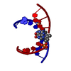
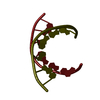
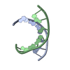
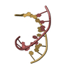
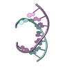
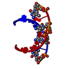
- PDB-9dna: CRYSTAL STRUCTURE ANALYSIS OF AN A-DNA FRAGMENT AT 1.8 ANGSTROMS ... -
+ Open data

- Basic information
| Entry | Database: PDB / ID: 9dna | ||||||||||||||||||
|---|---|---|---|---|---|---|---|---|---|---|---|---|---|---|---|---|---|---|---|
| Title | CRYSTAL STRUCTURE ANALYSIS OF AN A-DNA FRAGMENT AT 1.8 ANGSTROMS RESOLUTION. D(GCCCGGGC) | ||||||||||||||||||
 Components Components | DNA (5'-D(* Keywords KeywordsDNA / A-DNA / DOUBLE HELIX | Function / homology | DNA |  Function and homology information Function and homology informationMethod |  X-RAY DIFFRACTION / Resolution: 1.8 Å X-RAY DIFFRACTION / Resolution: 1.8 Å  Authors AuthorsHeinemann, U. |  CitationJournal: Nucleic Acids Res. / Year: 1987 CitationJournal: Nucleic Acids Res. / Year: 1987Title: Crystal structure analysis of an A-DNA fragment at 1.8 A resolution: d(GCCCGGGC). Authors: Heinemann, U. / Lauble, H. / Frank, R. / Blocker, H. History |
|
- Structure visualization
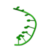
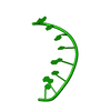
| Structure viewer | Molecule: MolmilJmol/JSmol |
|---|
- Downloads & links
-Download
| PDBx/mmCIF format | 9dna.cif.gz | 12.9 KB | Display | PDBx/mmCIF format |
|---|---|---|---|---|
| PDB format | pdb9dna.ent.gz | 7.7 KB | Display | PDB format |
| PDBx/mmJSON format | 9dna.json.gz | Tree view | PDBx/mmJSON format | |
| Others |  Other downloads Other downloads |
-Validation report
| Arichive directory | https://data.pdbj.org/pub/pdb/validation_reports/dn/9dnaftp://data.pdbj.org/pub/pdb/validation_reports/dn/9dna | HTTPS FTP |
|---|
-Related structure data
| Similar structure data |
|---|


-Links
 PDBj
PDBj

- Assembly
| Deposited unit | 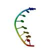
| ||||||||
|---|---|---|---|---|---|---|---|---|---|
| 1 | 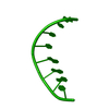
| ||||||||
| Unit cell |
| ||||||||
| Atom site foot note | 1: THE DEOXYRIBOSE PORTION OF RESIDUE C A 8 ADOPTS A SUGAR PUCKER IN THE C3(PRIME) -EXO DOMAIN. THIS IS UNUSUAL FOR THE A-DNA CONFORMATION. |
-Components
| #1: DNA chain | Mass: 2428.593 Da / Num. of mol.: 1 / Source method: obtained synthetically |
|---|---|
| #2: Water | ChemComp-HOH /  Mass: 18.015 Da / Num. of mol.: 34 / Source method: isolated from a natural source / Formula: H2O Mass: 18.015 Da / Num. of mol.: 34 / Source method: isolated from a natural source / Formula: H2O |
| Compound details | THE DEOXYRIBOSE PORTION OF RESIDUE C A 8 ADOPTS A SUGAR PUCKER IN THE C3(PRIME) -EXO DOMAIN. THIS ...THE DEOXYRIBOS |
-Experimental details
-Experiment
| Experiment | Method: X-RAY DIFFRACTION |
|---|
- Sample preparation
| Crystal | Density Matthews: 2.37 Å3/Da / Density % sol: 48.09 % | ||||||||||||||||||||||||||||||||||||||||||||||||||||||||
|---|---|---|---|---|---|---|---|---|---|---|---|---|---|---|---|---|---|---|---|---|---|---|---|---|---|---|---|---|---|---|---|---|---|---|---|---|---|---|---|---|---|---|---|---|---|---|---|---|---|---|---|---|---|---|---|---|---|
| Crystal grow | Method: microdialysis / Details: MICRODIALYSIS / Temp details: ROOM TEMPERATURE | ||||||||||||||||||||||||||||||||||||||||||||||||||||||||
| Components of the solutions |
| ||||||||||||||||||||||||||||||||||||||||||||||||||||||||
| Crystal grow | *PLUS pH: 7.5 | ||||||||||||||||||||||||||||||||||||||||||||||||||||||||
| Components of the solutions | *PLUS
|
-Data collection
| Diffraction | Ambient temp details: ROOM TEMPERATURE |
|---|---|
| Diffraction source | Source: SEALED TUBE / Wavelength: 1.5418 |
| Detector | Type: STOE / Detector: DIFFRACTOMETER |
| Radiation | Scattering type: x-ray |
| Radiation wavelength | Wavelength: 1.5418 Å / Relative weight: 1 |
| Reflection | Highest resolution: 1.8 Å / Num. all: 2599 / Num. obs: 1553 / Observed criterion σ(I): 1 |
| Reflection | *PLUS Observed criterion σ(I): 1 / Rmerge(I) obs: 0.027 / Num. measured all: 2599 |
- Processing
| Software | Name: NUCLSQ / Classification: refinement | |||||||||||||||||||||||||||||||||||||||||||||||||||||||||||||||
|---|---|---|---|---|---|---|---|---|---|---|---|---|---|---|---|---|---|---|---|---|---|---|---|---|---|---|---|---|---|---|---|---|---|---|---|---|---|---|---|---|---|---|---|---|---|---|---|---|---|---|---|---|---|---|---|---|---|---|---|---|---|---|---|---|
| Refinement | Resolution: 1.8→6 Å / σ(F): 3 /
| |||||||||||||||||||||||||||||||||||||||||||||||||||||||||||||||
| Refine Biso |
| |||||||||||||||||||||||||||||||||||||||||||||||||||||||||||||||
| Refinement step | Cycle: LAST / Resolution: 1.8→6 Å
| |||||||||||||||||||||||||||||||||||||||||||||||||||||||||||||||
| Refine LS restraints |
| |||||||||||||||||||||||||||||||||||||||||||||||||||||||||||||||
| Refinement | *PLUS Highest resolution: 1.8 Å / Lowest resolution: 6 Å / Num. reflection obs: 1359 / σ(F): 3 / Rfactor obs: 0.171 | |||||||||||||||||||||||||||||||||||||||||||||||||||||||||||||||
| Solvent computation | *PLUS | |||||||||||||||||||||||||||||||||||||||||||||||||||||||||||||||
| Displacement parameters | *PLUS | |||||||||||||||||||||||||||||||||||||||||||||||||||||||||||||||
| Refine LS restraints | *PLUS
|
