 Movie
Movie Controller

[English] 日本語
 Yorodumi
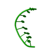
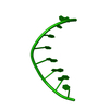
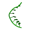
- PDB-1d26: EFFECT OF A SINGLE 3'-METHYLENE PHOSPHONATE LINKAGE ON THE CONFOR... -
+ Open data

- Basic information
| Entry | Database: PDB / ID: 1d26 | ||||||||||||||||||
|---|---|---|---|---|---|---|---|---|---|---|---|---|---|---|---|---|---|---|---|
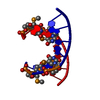
| Title | EFFECT OF A SINGLE 3'-METHYLENE PHOSPHONATE LINKAGE ON THE CONFORMATION OF AN A-DNA OCTAMER DOUBLE HELIX | ||||||||||||||||||
 Components Components | DNA (5'-D(* Keywords KeywordsDNA / A-DNA / DOUBLE HELIX / MODIFIED | Function / homology | DNA |  Function and homology information Function and homology informationMethod |  X-RAY DIFFRACTION / SYNCHROTRON / Resolution: 2.12 Å X-RAY DIFFRACTION / SYNCHROTRON / Resolution: 2.12 Å  Authors AuthorsHeinemann, U. |  CitationJournal: Nucleic Acids Res. / Year: 1991 CitationJournal: Nucleic Acids Res. / Year: 1991Title: Effect of a single 3'-methylene phosphonate linkage on the conformation of an A-DNA octamer double helix. Authors: Heinemann, U. / Rudolph, L.N. / Alings, C. / Morr, M. / Heikens, W. / Frank, R. / Blocker, H. #1: Journal: Structure and Methods. DNA and RNA / Year: 1990Title: Structural Features of G/C-Rich DNA Going A or B Authors: Heinemann, U. / Alings, C. / Lauble, H. #2: Journal: Nucleosides and Nucleotides / Year: 1990Title: X-Ray Studies of DNA Aiming at Elucidating the Sequence-Structure Code Authors: Heinemann, U. / Alings, C. / Lauble, H. #3: Journal: Nucleosides and Nucleotides / Year: 1988Title: X-Ray Crystallographic Studies of the A-Form of DNA Authors: Heinemann, U. / Lauble, H. / Frank, R. / Bloecker, H. #4: Journal: Nucleic Acids Res. / Year: 1987Title: Crystal Structure Analysis of an A-DNA Fragment at 1.8 Angstroms Resolution: d(GCCCGGGC) Authors: Heinemann, U. / Lauble, H. / Frank, R. / Bloecker, H. History |
|
- Structure visualization
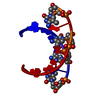
| Structure viewer | Molecule: MolmilJmol/JSmol |
|---|
- Downloads & links
-Download
| PDBx/mmCIF format | 1d26.cif.gz | 14 KB | Display | PDBx/mmCIF format |
|---|---|---|---|---|
| PDB format | pdb1d26.ent.gz | 8.3 KB | Display | PDB format |
| PDBx/mmJSON format | 1d26.json.gz | Tree view | PDBx/mmJSON format | |
| Others |  Other downloads Other downloads |
-Validation report
| Arichive directory | https://data.pdbj.org/pub/pdb/validation_reports/d2/1d26ftp://data.pdbj.org/pub/pdb/validation_reports/d2/1d26 | HTTPS FTP |
|---|
-Related structure data
| Similar structure data |
|---|


-Links
 PDBj
PDBj

- Assembly
| Deposited unit | 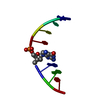
| ||||||||
|---|---|---|---|---|---|---|---|---|---|
| 1 | 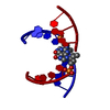
| ||||||||
| Unit cell |
|
-Components
| #1: DNA chain | Mass: 2426.620 Da / Num. of mol.: 1 Source method: isolated from a genetically manipulated source |
|---|---|
| #2: Water | ChemComp-HOH /  Mass: 18.015 Da / Num. of mol.: 29 / Source method: isolated from a natural source / Formula: H2O Mass: 18.015 Da / Num. of mol.: 29 / Source method: isolated from a natural source / Formula: H2O |
-Experimental details
-Experiment
| Experiment | Method: X-RAY DIFFRACTION |
|---|
- Sample preparation
| Crystal | Density Matthews: 2.37 Å3/Da / Density % sol: 48.06 % | ||||||||||||||||||||||||||||||||||||||||||
|---|---|---|---|---|---|---|---|---|---|---|---|---|---|---|---|---|---|---|---|---|---|---|---|---|---|---|---|---|---|---|---|---|---|---|---|---|---|---|---|---|---|---|---|
| Crystal grow | Temperature: 277 K / Method: microdialysis / pH: 7 / Details: pH 7.00, MICRODIALYSIS, temperature 277.00K | ||||||||||||||||||||||||||||||||||||||||||
| Components of the solutions |
| ||||||||||||||||||||||||||||||||||||||||||
| Crystal grow | *PLUS Temperature: 4 ℃ / pH: 7 | ||||||||||||||||||||||||||||||||||||||||||
| Components of the solutions | *PLUS
|
-Data collection
| Diffraction source | Source: SYNCHROTRON / Site: EMBL/DESY, HAMBURG  / Beamline: X31 / Beamline: X31 |
|---|---|
| Detector | Type: ARNDT-WONACOTT / Detector: OSCILLATION CAMERA |
| Radiation | Scattering type: x-ray |
| Radiation wavelength | Relative weight: 1 |
| Reflection | Resolution: 2.12→6 Å / Num. obs: 1303 / Observed criterion σ(F): 1 |
- Processing
| Software | Name: NUCLSQ / Classification: refinement | ||||||||||||||||||||||||||||||||||||||||||||||||||||||||||||||||||||||||||||||||||||
|---|---|---|---|---|---|---|---|---|---|---|---|---|---|---|---|---|---|---|---|---|---|---|---|---|---|---|---|---|---|---|---|---|---|---|---|---|---|---|---|---|---|---|---|---|---|---|---|---|---|---|---|---|---|---|---|---|---|---|---|---|---|---|---|---|---|---|---|---|---|---|---|---|---|---|---|---|---|---|---|---|---|---|---|---|---|
| Refinement | Resolution: 2.12→6 Å / σ(F): 3 /
| ||||||||||||||||||||||||||||||||||||||||||||||||||||||||||||||||||||||||||||||||||||
| Refine Biso |
| ||||||||||||||||||||||||||||||||||||||||||||||||||||||||||||||||||||||||||||||||||||
| Refinement step | Cycle: LAST / Resolution: 2.12→6 Å
| ||||||||||||||||||||||||||||||||||||||||||||||||||||||||||||||||||||||||||||||||||||
| Refine LS restraints |
| ||||||||||||||||||||||||||||||||||||||||||||||||||||||||||||||||||||||||||||||||||||
| Refinement | *PLUS Highest resolution: 2.12 Å / Lowest resolution: 6 Å / Num. reflection obs: 1197 / σ(I): 3 / Rfactor obs: 0.16 | ||||||||||||||||||||||||||||||||||||||||||||||||||||||||||||||||||||||||||||||||||||
| Solvent computation | *PLUS | ||||||||||||||||||||||||||||||||||||||||||||||||||||||||||||||||||||||||||||||||||||
| Displacement parameters | *PLUS | ||||||||||||||||||||||||||||||||||||||||||||||||||||||||||||||||||||||||||||||||||||
| Refine LS restraints | *PLUS
|
