 Movie
Movie Controller

[English] 日本語
 Yorodumi
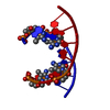
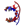
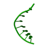
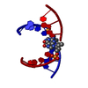
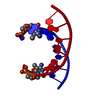
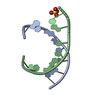
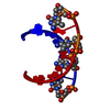
- PDB-1vt5: THE CRYSTAL STRUCTURE OF D(CCCCGGGG): A NEW A-FORM VARIANT WITH A... -
+ Open data

- Basic information
| Entry | Database: PDB / ID: 1vt5 | ||||||||||||||||||
|---|---|---|---|---|---|---|---|---|---|---|---|---|---|---|---|---|---|---|---|
| Title | THE CRYSTAL STRUCTURE OF D(CCCCGGGG): A NEW A-FORM VARIANT WITH AN EXTENDED BACKBONE CONFORMATION | ||||||||||||||||||
 Components Components | DNA (5'-D(* Keywords KeywordsDNA / A-DNA / DOUBLE HELIX | Function / homology | DNA |  Function and homology information Function and homology informationMethod |  X-RAY DIFFRACTION / Resolution: 2.25 Å X-RAY DIFFRACTION / Resolution: 2.25 Å  Authors AuthorsHaran, T.E. / Shakked, Z. / Wang, A.H.-J. / Rich, A. |  CitationJournal: J.Biomol.Struct.Dyn. / Year: 1987 CitationJournal: J.Biomol.Struct.Dyn. / Year: 1987Title: The crystal structure of d(CCCCGGGG): a new A-form variant with an extended backbone conformation. Authors: Haran, T.E. / Shakked, Z. / Wang, A.H. / Rich, A. #1: Journal: Proc.Natl.Acad.Sci.USA / Year: 1982Title: Molecular structure of the octamer d(G-G-C-C-G-G-C-C): modified A-DNA. Authors: Wang, A.H. / Fujii, S. / van Boom, J.H. / Rich, A. History |
|
- Structure visualization
| Structure viewer | Molecule: MolmilJmol/JSmol |
|---|
- Downloads & links
-Download
| PDBx/mmCIF format | 1vt5.cif.gz | 11.2 KB | Display | PDBx/mmCIF format |
|---|---|---|---|---|
| PDB format | pdb1vt5.ent.gz | 6.9 KB | Display | PDB format |
| PDBx/mmJSON format | 1vt5.json.gz | Tree view | PDBx/mmJSON format | |
| Others |  Other downloads Other downloads |
-Validation report
| Arichive directory | https://data.pdbj.org/pub/pdb/validation_reports/vt/1vt5ftp://data.pdbj.org/pub/pdb/validation_reports/vt/1vt5 | HTTPS FTP |
|---|
-Related structure data
| Similar structure data |
|---|


-Links
 PDBj
PDBj

- Assembly
| Deposited unit | 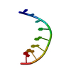
| ||||||||
|---|---|---|---|---|---|---|---|---|---|
| 1 | 
| ||||||||
| Unit cell |
|
-Components
| #1: DNA chain | Mass: 2428.593 Da / Num. of mol.: 1 / Source method: obtained synthetically / Details: SYNTHESIZED DNA |
|---|
-Experimental details
-Experiment
| Experiment | Method: X-RAY DIFFRACTION |
|---|
- Sample preparation
| Crystal | Density Matthews: 2.4 Å3/Da / Density % sol: 48.81 % | ||||||||||||||||||||||||||||||||||||||||||||||||||||||||
|---|---|---|---|---|---|---|---|---|---|---|---|---|---|---|---|---|---|---|---|---|---|---|---|---|---|---|---|---|---|---|---|---|---|---|---|---|---|---|---|---|---|---|---|---|---|---|---|---|---|---|---|---|---|---|---|---|---|
| Crystal grow | pH: 7 / Details: MPD, Sodium Cacodylate, spermine, MgCl2, pH 7.00 | ||||||||||||||||||||||||||||||||||||||||||||||||||||||||
| Components of the solutions |
|
-Data collection
| Diffraction |
| |||||||||||||||
|---|---|---|---|---|---|---|---|---|---|---|---|---|---|---|---|---|
| Diffraction source |
| |||||||||||||||
| Detector |
| |||||||||||||||
| Radiation |
| |||||||||||||||
| Radiation wavelength |
| |||||||||||||||
| Reflection | Highest resolution: 2.25 Å |
- Processing
| Software | Name: CORELS / Classification: refinement | ||||||||||||
|---|---|---|---|---|---|---|---|---|---|---|---|---|---|
| Refinement | Resolution: 2.25→8 Å Details: REFERENCE 2 REPORTED A CELL OF 42.10, 42.10, 25.10 WITH SPACE GROUP OF P 43 21 2. THIS HAS SINCE BEEN CHANGED TO THE PRESENT CELL.
| ||||||||||||
| Refine Biso | Class: ALL ATOMS / Details: TR / Treatment: isotropic | ||||||||||||
| Refinement step | Cycle: LAST / Resolution: 2.25→8 Å
|
