 Movie
Movie Controller

[English] 日本語
 Yorodumi
- PDB-9dn7: CryoEM structures of yeast cytoplasmic dynein in the presence of ... -
+ Open data

- Basic information
| Entry | Database: PDB / ID: 9dn7 | ||||||
|---|---|---|---|---|---|---|---|
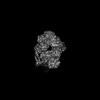
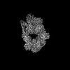
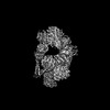
| Title | CryoEM structures of yeast cytoplasmic dynein in the presence of ATP and Lis1. | ||||||
 Components Components |
| ||||||
 Keywords Keywords | MOTOR PROTEIN / Enzyme / AAA protein | ||||||
| Function / homology |  Function and homology information Function and homology informationmicrotubule sliding / karyogamy / microtubule organizing center organization / nuclear migration along microtubule / astral microtubule / establishment of mitotic spindle localization / microtubule plus-end binding / spindle pole body / vesicle transport along microtubule / minus-end-directed microtubule motor activity ...microtubule sliding / karyogamy / microtubule organizing center organization / nuclear migration along microtubule / astral microtubule / establishment of mitotic spindle localization / microtubule plus-end binding / spindle pole body / vesicle transport along microtubule / minus-end-directed microtubule motor activity / microtubule associated complex / dynein light intermediate chain binding / cytoplasmic dynein complex / nuclear migration / Ubiquitin-Mediated Degradation of Phosphorylated Cdc25A / establishment of mitotic spindle orientation / dynein intermediate chain binding / Antigen processing: Ubiquitination & Proteasome degradation / mitotic sister chromatid segregation / dynein complex binding / cytoplasmic microtubule / cytoplasmic microtubule organization / Neutrophil degranulation / mitotic spindle organization / kinetochore / spindle pole / nuclear envelope / cell cortex / cell division / ATP binding / identical protein binding / nucleus / cytoplasm Similarity search - Function | ||||||
| Biological species |  | ||||||
| Method | ELECTRON MICROSCOPY / single particle reconstruction / cryo EM / Resolution: 3.25 Å | ||||||
 Authors Authors | Kendrick, A.A. / Leschziner, A.E. | ||||||
| Funding support |  United States, 1items United States, 1items
| ||||||
 Citation Citation | Journal: Nat Struct Mol Biol / Year: 2025 Title: Multiple steps of dynein activation by Lis1 visualized by cryo-EM. Authors: Agnieszka A Kendrick / Kendrick H V Nguyen / Wen Ma / Eva P Karasmanis / Rommie E Amaro / Samara L Reck-Peterson / Andres E Leschziner / Abstract: Cytoplasmic dynein-1 (dynein) is an essential molecular motor controlled in part by autoinhibition. Lis1, a key dynein regulator mutated in the neurodevelopmental disease lissencephaly, plays a role ...Cytoplasmic dynein-1 (dynein) is an essential molecular motor controlled in part by autoinhibition. Lis1, a key dynein regulator mutated in the neurodevelopmental disease lissencephaly, plays a role in dynein activation. We recently identified a structure of partially autoinhibited dynein bound to Lis1, which suggests an intermediate state in dynein's activation pathway. However, other structural information is needed to fully understand how Lis1 activates dynein. Here, we used cryo-EM and yeast dynein and Lis1 incubated with ATP at different time points to reveal conformations that we propose represent additional intermediate states in dynein's activation pathway. We solved 16 high-resolution structures, including 7 distinct dynein and dynein-Lis1 structures from the same sample. Our data support a model in which Lis1 relieves dynein autoinhibition by increasing its basal ATP hydrolysis rate and promoting conformations compatible with complex assembly and motility. Together, this analysis advances our understanding of dynein activation and the contribution of Lis1 to this process. | ||||||
| History |
|
- Structure visualization
| Structure viewer | Molecule: MolmilJmol/JSmol |
|---|
- Downloads & links
-Download
| PDBx/mmCIF format | 9dn7.cif.gz | 623.5 KB | Display | PDBx/mmCIF format |
|---|---|---|---|---|
| PDB format | pdb9dn7.ent.gz | 481.5 KB | Display | PDB format |
| PDBx/mmJSON format | 9dn7.json.gz | Tree view | PDBx/mmJSON format | |
| Others |  Other downloads Other downloads |
-Validation report
| Arichive directory | https://data.pdbj.org/pub/pdb/validation_reports/dn/9dn7ftp://data.pdbj.org/pub/pdb/validation_reports/dn/9dn7 | HTTPS FTP |
|---|
-Related structure data
| Related structure data |  47032MC  9di3C  9diuC  9dj7C  9djuC  9djyC  9djzC  9dk0C  9dkdC  9dkeC 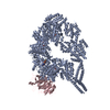 9dkhC  9dkjC 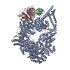 9dkmC 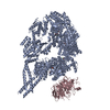 9dkxC  9dldC  9dleC  9dmwC  9dn5C 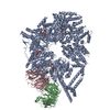 9dnbC C: citing same article ( M: map data used to model this data |
|---|---|
| Similar structure data |


-Links
 PDBj
PDBj


- Assembly
| Deposited unit | 
|
|---|---|
| 1 |
|
-Components
| #1: Protein | Mass: 331525.000 Da / Num. of mol.: 1 Source method: isolated from a genetically manipulated source Source: (gene. exp.) Gene: DYN1, DHC1, YKR054C / Production host: | ||||||||||
|---|---|---|---|---|---|---|---|---|---|---|---|
| #2: Protein | Mass: 57030.617 Da / Num. of mol.: 2 Source method: isolated from a genetically manipulated source Source: (gene. exp.) Gene: PAC1, LIS1, YOR269W / Production host: #3: Chemical | ChemComp-ATP / |   Mass: 507.181 Da / Num. of mol.: 1 / Source method: obtained synthetically / Formula: C10H16N5O13P3 / Feature type: SUBJECT OF INVESTIGATION / Comment: ATP, energy-carrying molecule*YM Mass: 507.181 Da / Num. of mol.: 1 / Source method: obtained synthetically / Formula: C10H16N5O13P3 / Feature type: SUBJECT OF INVESTIGATION / Comment: ATP, energy-carrying molecule*YM#4: Chemical |   Mass: 427.201 Da / Num. of mol.: 3 / Source method: obtained synthetically / Formula: C10H15N5O10P2 / Feature type: SUBJECT OF INVESTIGATION / Comment: ADP, energy-carrying molecule*YM Mass: 427.201 Da / Num. of mol.: 3 / Source method: obtained synthetically / Formula: C10H15N5O10P2 / Feature type: SUBJECT OF INVESTIGATION / Comment: ADP, energy-carrying molecule*YM#5: Chemical | ChemComp-MG / |   Mass: 24.305 Da / Num. of mol.: 1 / Source method: obtained synthetically / Formula: Mg / Feature type: SUBJECT OF INVESTIGATION Mass: 24.305 Da / Num. of mol.: 1 / Source method: obtained synthetically / Formula: Mg / Feature type: SUBJECT OF INVESTIGATIONHas ligand of interest | Y | Has protein modification | N | |
-Experimental details
-Experiment
| Experiment | Method: ELECTRON MICROSCOPY |
|---|---|
| EM experiment | Aggregation state: PARTICLE / 3D reconstruction method: single particle reconstruction |
- Sample preparation
| Component | Name: yeast dynein in the presence of ATP and PAC1 / Type: COMPLEX / Entity ID: #1-#2 / Source: RECOMBINANT |
|---|---|
| Source (natural) | Organism: |
| Source (recombinant) | Organism: |
| Buffer solution | pH: 8 Details: 50 mM TrisHCl [pH 8.0], 150 mM KOAc, 2 mM MgOAc, 1mM EGTA, 1 mM DTT |
| Specimen | Embedding applied: NO / Shadowing applied: NO / Staining applied: NO / Vitrification applied: YES |
| Vitrification | Instrument: FEI VITROBOT MARK II / Cryogen name: ETHANE / Humidity: 100 % / Chamber temperature: 277.15 K |
- Electron microscopy imaging
| Experimental equipment |  Model: Titan Krios / Image courtesy: FEI Company |
|---|---|
| Microscopy | Model: FEI TITAN KRIOS |
| Electron gun | Electron source:  FIELD EMISSION GUN / Accelerating voltage: 200 kV / Illumination mode: FLOOD BEAM FIELD EMISSION GUN / Accelerating voltage: 200 kV / Illumination mode: FLOOD BEAM |
| Electron lens | Mode: BRIGHT FIELD / Nominal defocus max: 2800 nm / Nominal defocus min: 750 nm / Cs: 2.7 mm |
| Image recording | Electron dose: 54 e/Å2 / Film or detector model: FEI FALCON IV (4k x 4k) |
- Processing
| EM software | Name: PHENIX / Category: model refinement | ||||||||||||||||||||||||
|---|---|---|---|---|---|---|---|---|---|---|---|---|---|---|---|---|---|---|---|---|---|---|---|---|---|
| CTF correction | Type: PHASE FLIPPING AND AMPLITUDE CORRECTION | ||||||||||||||||||||||||
| 3D reconstruction | Resolution: 3.25 Å / Resolution method: FSC 0.143 CUT-OFF / Num. of particles: 28077 / Symmetry type: POINT | ||||||||||||||||||||||||
| Refine LS restraints |
|
