 Movie
Movie Controller


+ Open data

- Basic information
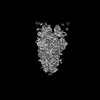
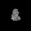
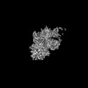
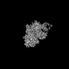
| Entry | Database: PDB / ID: 9df0 | |||||||||||||||||||||||||||||||||||||||||||||||||||||||||||||||||||||||||||||||||
|---|---|---|---|---|---|---|---|---|---|---|---|---|---|---|---|---|---|---|---|---|---|---|---|---|---|---|---|---|---|---|---|---|---|---|---|---|---|---|---|---|---|---|---|---|---|---|---|---|---|---|---|---|---|---|---|---|---|---|---|---|---|---|---|---|---|---|---|---|---|---|---|---|---|---|---|---|---|---|---|---|---|---|
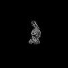
| Title | PDCoV S RBD bound to PD41 Fab (local refinement) | |||||||||||||||||||||||||||||||||||||||||||||||||||||||||||||||||||||||||||||||||
 Components Components |
| |||||||||||||||||||||||||||||||||||||||||||||||||||||||||||||||||||||||||||||||||
 Keywords Keywords | VIRAL PROTEIN / coronavirus / spike / antibody / Fab / antigen / PDCoV / PD41 / complex / Structural Genomics / Seattle Structural Genomics Center for Infectious Disease / SSGCID | |||||||||||||||||||||||||||||||||||||||||||||||||||||||||||||||||||||||||||||||||
| Function / homology |  Function and homology information Function and homology informationhost cell endoplasmic reticulum-Golgi intermediate compartment membrane / receptor-mediated virion attachment to host cell / endocytosis involved in viral entry into host cell / fusion of virus membrane with host plasma membrane / fusion of virus membrane with host endosome membrane / viral envelope / virion membrane / membrane Similarity search - Function | |||||||||||||||||||||||||||||||||||||||||||||||||||||||||||||||||||||||||||||||||
| Biological species |  Porcine deltacoronavirus Porcine deltacoronavirus | |||||||||||||||||||||||||||||||||||||||||||||||||||||||||||||||||||||||||||||||||
| Method | ELECTRON MICROSCOPY / single particle reconstruction / cryo EM / Resolution: 2.8 Å | |||||||||||||||||||||||||||||||||||||||||||||||||||||||||||||||||||||||||||||||||
 Authors Authors | Asarnow, D. / Rexhepaj, M. / Seattle Structural Genomics Center for Infectious Disease (SSGCID) / Veesler, D. | |||||||||||||||||||||||||||||||||||||||||||||||||||||||||||||||||||||||||||||||||
| Funding support |  United States, 6items United States, 6items
| |||||||||||||||||||||||||||||||||||||||||||||||||||||||||||||||||||||||||||||||||
 Citation Citation | Journal: Immunity / Year: 2024 Title: Isolation and escape mapping of broadly neutralizing antibodies against emerging delta-coronaviruses. Authors: Megi Rexhepaj / Daniel Asarnow / Lisa Perruzza / Young-Jun Park / Barbara Guarino / Mathew Mccallum / Katja Culap / Christian Saliba / Giada Leoni / Alessio Balmelli / Courtney N Yoshiyama / ...Authors: Megi Rexhepaj / Daniel Asarnow / Lisa Perruzza / Young-Jun Park / Barbara Guarino / Mathew Mccallum / Katja Culap / Christian Saliba / Giada Leoni / Alessio Balmelli / Courtney N Yoshiyama / Miles S Dickinson / Joel Quispe / Jack T Brown / M Alejandra Tortorici / Kaitlin R Sprouse / Ashley L Taylor / Davide Corti / Tyler N Starr / Fabio Benigni / David Veesler /  Abstract: Porcine delta-coronavirus (PDCoV) spillovers were recently detected in febrile children, underscoring the recurrent zoonoses of divergent CoVs. To date, no vaccines or specific therapeutics are ...Porcine delta-coronavirus (PDCoV) spillovers were recently detected in febrile children, underscoring the recurrent zoonoses of divergent CoVs. To date, no vaccines or specific therapeutics are approved for use in humans against PDCoV. To prepare for possible future PDCoV epidemics, we isolated PDCoV spike (S)-directed monoclonal antibodies (mAbs) from humanized mice and found that two, designated PD33 and PD41, broadly neutralized a panel of PDCoV variants. Cryoelectron microscopy (cryo-EM) structures of PD33 and PD41 in complex with the S receptor-binding domain (RBD) and ectodomain trimer revealed the epitopes recognized by these mAbs, rationalizing their broad inhibitory activity. We show that both mAbs competitively interfere with host aminopeptidase N binding to neutralize PDCoV and used deep-mutational scanning epitope mapping to associate RBD antigenic sites with mAb-mediated neutralization potency. Our results indicate a PD33-PD41 mAb cocktail may heighten the barrier to escape. PD33 and PD41 are candidates for clinical advancement against future PDCoV outbreaks. | |||||||||||||||||||||||||||||||||||||||||||||||||||||||||||||||||||||||||||||||||
| History |
|
- Structure visualization
| Structure viewer | Molecule: MolmilJmol/JSmol |
|---|
- Downloads & links
-Download
| PDBx/mmCIF format | 9df0.cif.gz | 119.6 KB | Display | PDBx/mmCIF format |
|---|---|---|---|---|
| PDB format | pdb9df0.ent.gz | 62 KB | Display | PDB format |
| PDBx/mmJSON format | 9df0.json.gz | Tree view | PDBx/mmJSON format | |
| Others |  Other downloads Other downloads |
-Validation report
| Arichive directory | https://data.pdbj.org/pub/pdb/validation_reports/df/9df0ftp://data.pdbj.org/pub/pdb/validation_reports/df/9df0 | HTTPS FTP |
|---|
-Related structure data
| Related structure data | 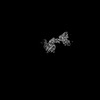 46805MC 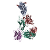 9b2cC 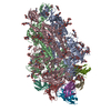 9dezC C: citing same article ( M: map data used to model this data |
|---|---|
| Similar structure data |

-Links
 PDBj
PDBj

- Assembly
| Deposited unit | 
|
|---|---|
| 1 |
|
-Components
| #1: Protein | Mass: 128934.805 Da / Num. of mol.: 1 Source method: isolated from a genetically manipulated source Source: (gene. exp.) Porcine deltacoronavirus / Variant: SD2018/300 / Cell line (production host): expi293F / Production host:  Homo sapiens (human) / References: UniProt: A0A6M5ICE2 Homo sapiens (human) / References: UniProt: A0A6M5ICE2 |
|---|---|
| #2: Antibody | Mass: 13020.510 Da / Num. of mol.: 1 Source method: isolated from a genetically manipulated source Source: (gene. exp.)  Cricetulus griseus (Chinese hamster) Cricetulus griseus (Chinese hamster) |
| #3: Antibody | Mass: 11603.645 Da / Num. of mol.: 1 Source method: isolated from a genetically manipulated source Source: (gene. exp.) Cricetulus griseus (Chinese hamster) |
| #4: Polysaccharide | beta-D-mannopyranose-(1-4)-2-acetamido-2-deoxy-beta-D-glucopyranose-(1-4)-2-acetamido-2-deoxy-beta- ...beta-D-mannopyranose-(1-4)-2-acetamido-2-deoxy-beta-D-glucopyranose-(1-4)-2-acetamido-2-deoxy-beta-D-glucopyranose Source method: isolated from a genetically manipulated source |
| #5: Sugar | ChemComp-NAG /   Type: D-saccharide, beta linking / Mass: 221.208 Da / Num. of mol.: 1 / Source method: obtained synthetically / Formula: C8H15NO6 Type: D-saccharide, beta linking / Mass: 221.208 Da / Num. of mol.: 1 / Source method: obtained synthetically / Formula: C8H15NO6 |
| Has ligand of interest | N |
| Has protein modification | Y |
-Experimental details
-Experiment
| Experiment | Method: ELECTRON MICROSCOPY |
|---|---|
| EM experiment | Aggregation state: PARTICLE / 3D reconstruction method: single particle reconstruction |
- Sample preparation
| Component | Name: PDCoV S RBD complex with PD41 Fab (local refinement) / Type: COMPLEX / Entity ID: #1-#3 / Source: RECOMBINANT |
|---|---|
| Molecular weight | Value: 0.04 MDa / Experimental value: NO |
| Source (natural) | Organism: Deltacoronavirus PDCoV/USA/Illinois121/2014 |
| Source (recombinant) | Organism: Homo sapiens (human) / Cell: expi293F |
| Buffer solution | pH: 8 |
| Specimen | Embedding applied: NO / Shadowing applied: NO / Staining applied: NO / Vitrification applied: YES |
| Specimen support | Grid material: GOLD / Grid mesh size: 300 divisions/in. / Grid type: UltrAuFoil R1.2/1.3 |
| Vitrification | Instrument: FEI VITROBOT MARK IV / Cryogen name: ETHANE / Humidity: 100 % / Chamber temperature: 277 K / Details: Blotting time 6 seconds, force -1 |
- Electron microscopy imaging
| Experimental equipment |  Model: Titan Krios / Image courtesy: FEI Company |
|---|---|
| Microscopy | Model: FEI TITAN KRIOS |
| Electron gun | Electron source:  FIELD EMISSION GUN / Accelerating voltage: 300 kV / Illumination mode: FLOOD BEAM FIELD EMISSION GUN / Accelerating voltage: 300 kV / Illumination mode: FLOOD BEAM |
| Electron lens | Mode: BRIGHT FIELD / Nominal magnification: 105000 X / Nominal defocus max: 1800 nm / Nominal defocus min: 500 nm / Cs: 2.7 mm / Alignment procedure: COMA FREE |
| Specimen holder | Cryogen: NITROGEN / Specimen holder model: FEI TITAN KRIOS AUTOGRID HOLDER |
| Image recording | Average exposure time: 4 sec. / Electron dose: 45 e/Å2 / Film or detector model: GATAN K3 BIOQUANTUM (6k x 4k) / Num. of grids imaged: 2 / Num. of real images: 19556 |
| EM imaging optics | Energyfilter name: GIF Bioquantum / Energyfilter slit width: 20 eV |
| Image scans | Sampling size: 5 µm / Width: 5760 / Height: 4092 |
- Processing
| EM software |
| ||||||||||||||||||||||||||||||||||||||||||||||||
|---|---|---|---|---|---|---|---|---|---|---|---|---|---|---|---|---|---|---|---|---|---|---|---|---|---|---|---|---|---|---|---|---|---|---|---|---|---|---|---|---|---|---|---|---|---|---|---|---|---|
| CTF correction | Type: PHASE FLIPPING AND AMPLITUDE CORRECTION | ||||||||||||||||||||||||||||||||||||||||||||||||
| Particle selection | Num. of particles selected: 5732849 | ||||||||||||||||||||||||||||||||||||||||||||||||
| Symmetry | Point symmetry: C1 (asymmetric) | ||||||||||||||||||||||||||||||||||||||||||||||||
| 3D reconstruction | Resolution: 2.8 Å / Resolution method: FSC 0.143 CUT-OFF / Num. of particles: 438204 / Algorithm: FOURIER SPACE / Num. of class averages: 1 / Symmetry type: POINT | ||||||||||||||||||||||||||||||||||||||||||||||||
| Atomic model building | Protocol: FLEXIBLE FIT / Space: REAL | ||||||||||||||||||||||||||||||||||||||||||||||||
| Atomic model building | 3D fitting-ID: 1
| ||||||||||||||||||||||||||||||||||||||||||||||||
| Refinement | Cross valid method: NONE Stereochemistry target values: GeoStd + Monomer Library + CDL v1.2 | ||||||||||||||||||||||||||||||||||||||||||||||||
| Displacement parameters | Biso mean: 45.25 Å2 | ||||||||||||||||||||||||||||||||||||||||||||||||
| Refine LS restraints |
|

