[English] 日本語
 Yorodumi
- PDB-9d69: Human excitatory amino acid transporter 3 (EAAT3) with bound L-Cy... -
+ Open data
- Basic information
| Entry | Database: PDB / ID: 9d69 | |||||||||
|---|---|---|---|---|---|---|---|---|---|---|
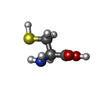
| Title | Human excitatory amino acid transporter 3 (EAAT3) with bound L-Cysteine in an intermediate outward facing state (slight upper position) | |||||||||
 Components Components | Excitatory amino acid transporter 3 | |||||||||
 Keywords Keywords | TRANSPORT PROTEIN / human EAAT3 / Cysteine / iOFS-upper | |||||||||
| Function / homology |  Function and homology information Function and homology informationD-aspartate transmembrane transport / regulation of protein targeting to membrane / D-aspartate transmembrane transporter activity / Defective SLC1A1 is implicated in schizophrenia 18 (SCZD18) and dicarboxylic aminoaciduria (DCBXA) / distal dendrite / cysteine transport / cysteine transmembrane transporter activity / neurotransmitter receptor transport to plasma membrane / high-affinity L-glutamate transmembrane transporter activity / glutamate:sodium symporter activity ...D-aspartate transmembrane transport / regulation of protein targeting to membrane / D-aspartate transmembrane transporter activity / Defective SLC1A1 is implicated in schizophrenia 18 (SCZD18) and dicarboxylic aminoaciduria (DCBXA) / distal dendrite / cysteine transport / cysteine transmembrane transporter activity / neurotransmitter receptor transport to plasma membrane / high-affinity L-glutamate transmembrane transporter activity / glutamate:sodium symporter activity / L-glutamate import / response to decreased oxygen levels / cellular response to mercury ion / : / L-glutamate transmembrane transporter activity / retina layer formation / L-glutamate transmembrane transport / glutathione biosynthetic process / D-aspartate import across plasma membrane / L-aspartate transmembrane transport / cellular response to ammonium ion / righting reflex / zinc ion transmembrane transport / L-aspartate transmembrane transporter activity / cellular response to bisphenol A / intracellular glutamate homeostasis / grooming behavior / L-aspartate import across plasma membrane / Glutamate Neurotransmitter Release Cycle / L-glutamate import across plasma membrane / proximal dendrite / monoatomic anion channel activity / transepithelial transport / apical dendrite / intracellular zinc ion homeostasis / blood vessel morphogenesis / cellular response to cocaine / chloride transmembrane transporter activity / motor neuron apoptotic process / response to anesthetic / response to morphine / glutamate receptor signaling pathway / G protein-coupled dopamine receptor signaling pathway / superoxide metabolic process / heart contraction / neurotransmitter transport / maintenance of blood-brain barrier / perisynaptic space / dopamine metabolic process / adult behavior / motor behavior / asymmetric synapse / conditioned place preference / glial cell projection / synaptic cleft / response to axon injury / behavioral fear response / positive regulation of heart rate / postsynaptic modulation of chemical synaptic transmission / transport across blood-brain barrier / monoatomic ion transport / neurogenesis / axon terminus / chloride transmembrane transport / response to amphetamine / dendritic shaft / cell periphery / locomotory behavior / synapse organization / brain development / Schaffer collateral - CA1 synapse / memory / long-term synaptic potentiation / recycling endosome membrane / cytokine-mediated signaling pathway / late endosome membrane / presynapse / cellular response to oxidative stress / early endosome membrane / perikaryon / gene expression / dendritic spine / chemical synaptic transmission / negative regulation of neuron apoptotic process / apical plasma membrane / membrane raft / response to xenobiotic stimulus / axon / neuronal cell body / dendrite / cell surface / extracellular exosome / metal ion binding / identical protein binding / membrane / plasma membrane Similarity search - Function | |||||||||
| Biological species |  Homo sapiens (human) Homo sapiens (human) | |||||||||
| Method | ELECTRON MICROSCOPY / single particle reconstruction / cryo EM / Resolution: 2.99 Å | |||||||||
 Authors Authors | Qiu, B. / Boudker, O. | |||||||||
| Funding support |  United States, 2items United States, 2items
| |||||||||
 Citation Citation |  Journal: bioRxiv / Year: 2024 Journal: bioRxiv / Year: 2024Title: Structural basis of the excitatory amino acid transporter 3 substrate recognition. Authors: Biao Qiu / Olga Boudker /  Abstract: Excitatory amino acid transporters (EAATs) reside on cell surfaces and uptake substrates, including L-glutamate, L-aspartate, and D-aspartate, using ion gradients. Among five EAATs, EAAT3 is the only ...Excitatory amino acid transporters (EAATs) reside on cell surfaces and uptake substrates, including L-glutamate, L-aspartate, and D-aspartate, using ion gradients. Among five EAATs, EAAT3 is the only isoform that can efficiently transport L-cysteine, a substrate for glutathione synthesis. Recent work suggests that EAAT3 also transports the oncometabolite R-2-hydroxyglutarate (R-2HG). Here, we examined the structural basis of substrate promiscuity by determining the cryo-EM structures of EAAT3 bound to different substrates. We found that L-cysteine binds to EAAT3 in thiolate form, and EAAT3 recognizes different substrates by fine-tuning local conformations of the coordinating residues. However, using purified human EAAT3, we could not observe R-2HG binding or transport. Imaging of EAAT3 bound to L-cysteine revealed several conformational states, including an outward-facing state with a semi-open gate and a disrupted sodium-binding site. These structures illustrate that the full gate closure, coupled with the binding of the last sodium ion, occurs after substrate binding. Furthermore, we observed that different substrates affect how the transporter distributes between a fully outward-facing conformation and intermediate occluded states on a path to the inward-facing conformation, suggesting that translocation rates are substrate-dependent. | |||||||||
| History |
|
- Structure visualization
| Structure viewer | Molecule:  Molmil Molmil Jmol/JSmol Jmol/JSmol |
|---|
- Downloads & links
- Download
| PDBx/mmCIF format |  9d69.cif.gz 9d69.cif.gz | 91.8 KB | Display |  PDBx/mmCIF format PDBx/mmCIF format |
|---|---|---|---|---|
| PDB format |  pdb9d69.ent.gz pdb9d69.ent.gz | 66.9 KB | Display |  PDB format PDB format |
| PDBx/mmJSON format |  9d69.json.gz 9d69.json.gz | Tree view |  PDBx/mmJSON format PDBx/mmJSON format | |
| Others |  Other downloads Other downloads |
-Validation report
| Summary document |  9d69_validation.pdf.gz 9d69_validation.pdf.gz | 1.5 MB | Display |  wwPDB validaton report wwPDB validaton report |
|---|---|---|---|---|
| Full document |  9d69_full_validation.pdf.gz 9d69_full_validation.pdf.gz | 1.5 MB | Display | |
| Data in XML |  9d69_validation.xml.gz 9d69_validation.xml.gz | 29.8 KB | Display | |
| Data in CIF |  9d69_validation.cif.gz 9d69_validation.cif.gz | 41.5 KB | Display | |
| Arichive directory |  https://data.pdbj.org/pub/pdb/validation_reports/d6/9d69 https://data.pdbj.org/pub/pdb/validation_reports/d6/9d69 ftp://data.pdbj.org/pub/pdb/validation_reports/d6/9d69 ftp://data.pdbj.org/pub/pdb/validation_reports/d6/9d69 | HTTPS FTP |
-Related structure data
| Related structure data |  46590MC  9d66C  9d67C  9d68C  9d6aC M: map data used to model this data C: citing same article ( |
|---|---|
| Similar structure data | Similarity search - Function & homology  F&H Search F&H Search |
- Links
Links
- Assembly
| Deposited unit | 
|
|---|---|
| 1 |
|
- Components
| #1: Protein | Mass: 57120.863 Da / Num. of mol.: 1 Source method: isolated from a genetically manipulated source Source: (gene. exp.)  Homo sapiens (human) / Gene: SLC1A1, EAAC1, EAAT3 / Production host: Homo sapiens (human) / Gene: SLC1A1, EAAC1, EAAT3 / Production host:  Homo sapiens (human) / References: UniProt: P43005 Homo sapiens (human) / References: UniProt: P43005 | ||||||
|---|---|---|---|---|---|---|---|
| #2: Chemical | ChemComp-CYS / | ||||||
| #3: Chemical | | #4: Chemical | ChemComp-HG / | Has ligand of interest | Y | Has protein modification | N | |
-Experimental details
-Experiment
| Experiment | Method: ELECTRON MICROSCOPY |
|---|---|
| EM experiment | Aggregation state: PARTICLE / 3D reconstruction method: single particle reconstruction |
- Sample preparation
| Component | Name: EAAT3 monomer with L-Cys bound at iOFS / Type: COMPLEX / Entity ID: #1 / Source: RECOMBINANT |
|---|---|
| Molecular weight | Experimental value: NO |
| Source (natural) | Organism:  Homo sapiens (human) Homo sapiens (human) |
| Source (recombinant) | Organism:  Homo sapiens (human) Homo sapiens (human) |
| Buffer solution | pH: 7.4 Details: 20 mM Hepes-Tris pH 7.4, 200 mM NaCl and 0.01% GDN, 10 mM L-Cysteine |
| Specimen | Conc.: 5 mg/ml / Embedding applied: NO / Shadowing applied: NO / Staining applied: NO / Vitrification applied: YES / Details: This sample was monodisperse. |
| Specimen support | Grid material: GOLD / Grid mesh size: 300 divisions/in. / Grid type: Quantifoil R1.2/1.3 |
| Vitrification | Instrument: FEI VITROBOT MARK IV / Cryogen name: ETHANE / Humidity: 100 % / Chamber temperature: 277 K |
- Electron microscopy imaging
| Microscopy | Model: FEI/PHILIPS CM300FEG/T |
|---|---|
| Electron gun | Electron source:  FIELD EMISSION GUN / Accelerating voltage: 300 kV / Illumination mode: FLOOD BEAM FIELD EMISSION GUN / Accelerating voltage: 300 kV / Illumination mode: FLOOD BEAM |
| Electron lens | Mode: BRIGHT FIELD / Nominal defocus max: 2400 nm / Nominal defocus min: 800 nm |
| Image recording | Electron dose: 58.25 e/Å2 / Film or detector model: GATAN K3 (6k x 4k) |
| EM imaging optics | Energyfilter slit width: 20 eV |
- Processing
| EM software |
| ||||||||||||||||||||||||||||||||||||
|---|---|---|---|---|---|---|---|---|---|---|---|---|---|---|---|---|---|---|---|---|---|---|---|---|---|---|---|---|---|---|---|---|---|---|---|---|---|
| CTF correction | Type: PHASE FLIPPING AND AMPLITUDE CORRECTION | ||||||||||||||||||||||||||||||||||||
| Particle selection | Num. of particles selected: 4180263 Details: This is the number of trimer particle from the image | ||||||||||||||||||||||||||||||||||||
| Symmetry | Point symmetry: C1 (asymmetric) | ||||||||||||||||||||||||||||||||||||
| 3D reconstruction | Resolution: 2.99 Å / Resolution method: FSC 0.143 CUT-OFF / Num. of particles: 60670 Details: This is number of monomer from C3 expanded particles. Num. of class averages: 1 / Symmetry type: POINT | ||||||||||||||||||||||||||||||||||||
| Atomic model building | B value: 59.17 / Protocol: OTHER | ||||||||||||||||||||||||||||||||||||
| Atomic model building | PDB-ID: 8CV3 Accession code: 8CV3 / Source name: PDB / Type: experimental model |
 Movie
Movie Controller


 PDBj
PDBj


