 Movie
Movie Controller


+ Open data

- Basic information
| Entry | Database: PDB / ID: 9d4z | ||||||||||||||||||||||||
|---|---|---|---|---|---|---|---|---|---|---|---|---|---|---|---|---|---|---|---|---|---|---|---|---|---|
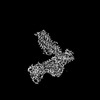
| Title | CryoEM structure of PAR1 with endogenous tethered ligand | ||||||||||||||||||||||||
 Components Components |
| ||||||||||||||||||||||||
 Keywords Keywords | IMMUNE SYSTEM / PAR2 / endogenous ligand. | ||||||||||||||||||||||||
| Function / homology |  Function and homology information Function and homology informationnegative regulation of renin secretion into blood stream / dendritic cell homeostasis / trans-synaptic signaling by endocannabinoid, modulating synaptic transmission / platelet dense tubular network / establishment of synaptic specificity at neuromuscular junction / thrombin-activated receptor activity / connective tissue replacement involved in inflammatory response wound healing / regulation of interleukin-1 beta production / platelet dense granule organization / cell-cell junction maintenance ...negative regulation of renin secretion into blood stream / dendritic cell homeostasis / trans-synaptic signaling by endocannabinoid, modulating synaptic transmission / platelet dense tubular network / establishment of synaptic specificity at neuromuscular junction / thrombin-activated receptor activity / connective tissue replacement involved in inflammatory response wound healing / regulation of interleukin-1 beta production / platelet dense granule organization / cell-cell junction maintenance / positive regulation of smooth muscle contraction / positive regulation of calcium ion transport / thrombin-activated receptor signaling pathway / negative regulation of glomerular filtration / regulation of blood coagulation / positive regulation of Rho protein signal transduction / positive regulation of collagen biosynthetic process / positive regulation of blood coagulation / positive regulation of vasoconstriction / anatomical structure morphogenesis / : / G-protein alpha-subunit binding / release of sequestered calcium ion into cytosol / homeostasis of number of cells within a tissue / positive regulation of release of sequestered calcium ion into cytosol / positive regulation of interleukin-8 production / Peptide ligand-binding receptors / positive regulation of receptor signaling pathway via JAK-STAT / neuromuscular junction / platelet activation / response to wounding / caveola / regulation of synaptic plasticity / positive regulation of interleukin-6 production / G protein-coupled receptor activity / Olfactory Signaling Pathway / Activation of the phototransduction cascade / G protein-coupled acetylcholine receptor signaling pathway / G beta:gamma signalling through PLC beta / Presynaptic function of Kainate receptors / Thromboxane signalling through TP receptor / Activation of G protein gated Potassium channels / Inhibition of voltage gated Ca2+ channels via Gbeta/gamma subunits / G-protein activation / Glucagon signaling in metabolic regulation / G beta:gamma signalling through CDC42 / Prostacyclin signalling through prostacyclin receptor / Synthesis, secretion, and inactivation of Glucagon-like Peptide-1 (GLP-1) / G beta:gamma signalling through BTK / photoreceptor disc membrane / ADP signalling through P2Y purinoceptor 12 / late endosome / Glucagon-type ligand receptors / Sensory perception of sweet, bitter, and umami (glutamate) taste / Adrenaline,noradrenaline inhibits insulin secretion / Vasopressin regulates renal water homeostasis via Aquaporins / Glucagon-like Peptide-1 (GLP1) regulates insulin secretion / G alpha (z) signalling events / cellular response to catecholamine stimulus / ADP signalling through P2Y purinoceptor 1 / G beta:gamma signalling through PI3Kgamma / ADORA2B mediated anti-inflammatory cytokines production / adenylate cyclase-activating dopamine receptor signaling pathway / Cooperation of PDCL (PhLP1) and TRiC/CCT in G-protein beta folding / GPER1 signaling / cellular response to prostaglandin E stimulus / heterotrimeric G-protein complex / G alpha (12/13) signalling events / Inactivation, recovery and regulation of the phototransduction cascade / G-protein beta-subunit binding / extracellular vesicle / sensory perception of taste / Thrombin signalling through proteinase activated receptors (PARs) / signaling receptor complex adaptor activity / positive regulation of cytosolic calcium ion concentration / retina development in camera-type eye / fibroblast proliferation / GTPase binding / Ca2+ pathway / response to lipopolysaccharide / High laminar flow shear stress activates signaling by PIEZO1 and PECAM1:CDH5:KDR in endothelial cells / G alpha (i) signalling events / G alpha (s) signalling events / G alpha (q) signalling events / phospholipase C-activating G protein-coupled receptor signaling pathway / negative regulation of neuron apoptotic process / Ras protein signal transduction / early endosome / positive regulation of MAPK cascade / positive regulation of canonical NF-kappaB signal transduction / positive regulation of ERK1 and ERK2 cascade / postsynaptic membrane / positive regulation of phosphatidylinositol 3-kinase/protein kinase B signal transduction / Extra-nuclear estrogen signaling / cell population proliferation / positive regulation of cell migration / positive regulation of apoptotic process / G protein-coupled receptor signaling pathway / inflammatory response / signaling receptor binding Similarity search - Function | ||||||||||||||||||||||||
| Biological species |  Homo sapiens (human) Homo sapiens (human) | ||||||||||||||||||||||||
| Method | ELECTRON MICROSCOPY / single particle reconstruction / cryo EM / Resolution: 2.74 Å | ||||||||||||||||||||||||
 Authors Authors | Lyu, X. / Lyu, Z. / McGrath, A.P. / Kang, Y. | ||||||||||||||||||||||||
| Funding support | 1items
| ||||||||||||||||||||||||
 Citation Citation | Journal: Nat Commun / Year: 2025 Title: Structural basis for the activation of proteinase-activated receptors PAR1 and PAR2. Authors: Zongyang Lyu / Xiaoxuan Lyu / Andrey G Malyutin / Guliang Xia / Daniel Carney / Vinicius M Alves / Matthew Falk / Nidhi Arora / Hua Zou / Aaron P McGrath / Yanyong Kang /  Abstract: Members of the proteinase-activated receptor (PAR) subfamily of G protein-coupled receptors (GPCRs) play critical roles in processes like hemostasis, thrombosis, development, wound healing, ...Members of the proteinase-activated receptor (PAR) subfamily of G protein-coupled receptors (GPCRs) play critical roles in processes like hemostasis, thrombosis, development, wound healing, inflammation, and cancer progression. Comprising PAR1-PAR4, these receptors are specifically activated by protease cleavage at their extracellular amino terminus, revealing a 'tethered ligand' that self-activates the receptor. This triggers complex intracellular signaling via G proteins and beta-arrestins, linking external protease signals to cellular functions. To date, direct structural visualization of these ligand-receptor complexes has been limited. Here, we present structural snapshots of activated PAR1 and PAR2 bound to their endogenous tethered ligands, revealing a shallow and constricted orthosteric binding pocket. Comparisons with antagonist-bound structures show minimal conformational changes in the TM6 helix and larger movements of TM7 upon activation. These findings reveal a common activation mechanism for PAR1 and PAR2, highlighting critical residues involved in ligand recognition. Additionally, the structure of PAR2 bound to a pathway selective antagonist, GB88, demonstrates how potent orthosteric engagement can be achieved by a small molecule mimicking the endogenous tethered ligand's interactions. | ||||||||||||||||||||||||
| History |
|
- Structure visualization
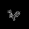
| Structure viewer | Molecule: MolmilJmol/JSmol |
|---|
- Downloads & links
-Download
| PDBx/mmCIF format | 9d4z.cif.gz | 224.2 KB | Display | PDBx/mmCIF format |
|---|---|---|---|---|
| PDB format | pdb9d4z.ent.gz | 170.8 KB | Display | PDB format |
| PDBx/mmJSON format | 9d4z.json.gz | Tree view | PDBx/mmJSON format | |
| Others |  Other downloads Other downloads |
-Validation report
| Arichive directory | https://data.pdbj.org/pub/pdb/validation_reports/d4/9d4zftp://data.pdbj.org/pub/pdb/validation_reports/d4/9d4z | HTTPS FTP |
|---|
-Related structure data
| Related structure data | 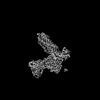 46571MC 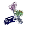 9d0aC 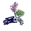 9e7rC M: map data used to model this data C: citing same article ( |
|---|---|
| Similar structure data |
-Links
 PDBj
PDBj


- Assembly
| Deposited unit | 
|
|---|---|
| 1 |
|
-Components
| #1: Protein | Mass: 37285.734 Da / Num. of mol.: 1 Source method: isolated from a genetically manipulated source Source: (gene. exp.) Homo sapiens (human) / Gene: GNB1 / Production host:   Spodoptera frugiperda (fall armyworm) / References: UniProt: P62873 Spodoptera frugiperda (fall armyworm) / References: UniProt: P62873 |
|---|---|
| #2: Protein | Mass: 6504.446 Da / Num. of mol.: 1 Source method: isolated from a genetically manipulated source Source: (gene. exp.) Homo sapiens (human) / Gene: GNG2 / Production host: Spodoptera frugiperda (fall armyworm) / References: UniProt: P59768 |
| #3: Protein | Mass: 43020.996 Da / Num. of mol.: 1 Source method: isolated from a genetically manipulated source Source: (gene. exp.) Homo sapiens (human) / Gene: F2R, CF2R, PAR1, TR / Production host: Spodoptera frugiperda (fall armyworm) / References: UniProt: P25116 |
| #4: Antibody | Mass: 26679.721 Da / Num. of mol.: 1 Source method: isolated from a genetically manipulated source Source: (gene. exp.) Spodoptera frugiperda (fall armyworm) |
| #5: Protein | Mass: 41972.578 Da / Num. of mol.: 1 Source method: isolated from a genetically manipulated source Source: (gene. exp.) Homo sapiens (human) / Production host: Spodoptera frugiperda (fall armyworm) |
| Has protein modification | Y |
-Experimental details
-Experiment
| Experiment | Method: ELECTRON MICROSCOPY |
|---|---|
| EM experiment | Aggregation state: PARTICLE / 3D reconstruction method: single particle reconstruction |
- Sample preparation
| Component | Name: Complex of PAR1 with G proteins / Type: COMPLEX / Entity ID: #3, #1-#2, #4-#5 / Source: MULTIPLE SOURCES |
|---|---|
| Molecular weight | Experimental value: NO |
| Source (natural) | Organism: Homo sapiens (human) |
| Source (recombinant) | Organism: Spodoptera frugiperda (fall armyworm) |
| Buffer solution | pH: 7 |
| Specimen | Embedding applied: NO / Shadowing applied: NO / Staining applied: NO / Vitrification applied: YES |
| Specimen support | Grid material: GOLD / Grid mesh size: 300 divisions/in. / Grid type: Quantifoil |
| Vitrification | Cryogen name: ETHANE |
- Electron microscopy imaging
| Microscopy | Model: TFS GLACIOS |
|---|---|
| Electron gun | Electron source:  FIELD EMISSION GUN / Accelerating voltage: 200 kV / Illumination mode: FLOOD BEAM FIELD EMISSION GUN / Accelerating voltage: 200 kV / Illumination mode: FLOOD BEAM |
| Electron lens | Mode: BRIGHT FIELD / Nominal defocus max: 2000 nm / Nominal defocus min: 800 nm |
| Image recording | Electron dose: 45 e/Å2 / Film or detector model: TFS FALCON 4i (4k x 4k) |
- Processing
| EM software | Name: PHENIX / Category: model refinement | ||||||||||||||||||||||||
|---|---|---|---|---|---|---|---|---|---|---|---|---|---|---|---|---|---|---|---|---|---|---|---|---|---|
| CTF correction | Type: PHASE FLIPPING AND AMPLITUDE CORRECTION | ||||||||||||||||||||||||
| 3D reconstruction | Resolution: 2.74 Å / Resolution method: FSC 0.143 CUT-OFF / Num. of particles: 1904551 / Symmetry type: POINT | ||||||||||||||||||||||||
| Refinement | Highest resolution: 2.74 Å Stereochemistry target values: REAL-SPACE (WEIGHTED MAP SUM AT ATOM CENTERS) | ||||||||||||||||||||||||
| Refine LS restraints |
|
