 Movie
Movie Controller


+ Open data

- Basic information
| Entry | 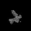 | |||||||||
|---|---|---|---|---|---|---|---|---|---|---|
| Title | CryoEM structure of PAR1 with endogenous tethered ligand | |||||||||
 Map data Map data | ||||||||||
 Sample Sample |
| |||||||||
 Keywords Keywords | PAR2 / endogenous ligand. / IMMUNE SYSTEM | |||||||||
| Function / homology |  Function and homology information Function and homology informationnegative regulation of renin secretion into blood stream / trans-synaptic signaling by endocannabinoid, modulating synaptic transmission / platelet dense tubular network / establishment of synaptic specificity at neuromuscular junction / thrombin-activated receptor activity / connective tissue replacement involved in inflammatory response wound healing / regulation of interleukin-1 beta production / cell-cell junction maintenance / platelet dense granule organization / positive regulation of smooth muscle contraction ...negative regulation of renin secretion into blood stream / trans-synaptic signaling by endocannabinoid, modulating synaptic transmission / platelet dense tubular network / establishment of synaptic specificity at neuromuscular junction / thrombin-activated receptor activity / connective tissue replacement involved in inflammatory response wound healing / regulation of interleukin-1 beta production / cell-cell junction maintenance / platelet dense granule organization / positive regulation of smooth muscle contraction / positive regulation of calcium ion transport / thrombin-activated receptor signaling pathway / negative regulation of glomerular filtration / regulation of blood coagulation / positive regulation of Rho protein signal transduction / positive regulation of collagen biosynthetic process / positive regulation of blood coagulation / positive regulation of vasoconstriction / anatomical structure morphogenesis / : / G-protein alpha-subunit binding / homeostasis of number of cells within a tissue / release of sequestered calcium ion into cytosol / positive regulation of release of sequestered calcium ion into cytosol / positive regulation of interleukin-8 production / Peptide ligand-binding receptors / positive regulation of receptor signaling pathway via JAK-STAT / neuromuscular junction / platelet activation / regulation of synaptic plasticity / response to wounding / caveola / positive regulation of interleukin-6 production / G protein-coupled receptor activity / Olfactory Signaling Pathway / Activation of the phototransduction cascade / G protein-coupled acetylcholine receptor signaling pathway / G beta:gamma signalling through PLC beta / Presynaptic function of Kainate receptors / Thromboxane signalling through TP receptor / Activation of G protein gated Potassium channels / Inhibition of voltage gated Ca2+ channels via Gbeta/gamma subunits / G-protein activation / Glucagon signaling in metabolic regulation / G beta:gamma signalling through CDC42 / Prostacyclin signalling through prostacyclin receptor / Synthesis, secretion, and inactivation of Glucagon-like Peptide-1 (GLP-1) / G beta:gamma signalling through BTK / photoreceptor disc membrane / ADP signalling through P2Y purinoceptor 12 / late endosome / Glucagon-type ligand receptors / Sensory perception of sweet, bitter, and umami (glutamate) taste / Adrenaline,noradrenaline inhibits insulin secretion / Vasopressin regulates renal water homeostasis via Aquaporins / Glucagon-like Peptide-1 (GLP1) regulates insulin secretion / G alpha (z) signalling events / cellular response to catecholamine stimulus / ADP signalling through P2Y purinoceptor 1 / G beta:gamma signalling through PI3Kgamma / ADORA2B mediated anti-inflammatory cytokines production / adenylate cyclase-activating dopamine receptor signaling pathway / Cooperation of PDCL (PhLP1) and TRiC/CCT in G-protein beta folding / GPER1 signaling / cellular response to prostaglandin E stimulus / heterotrimeric G-protein complex / Inactivation, recovery and regulation of the phototransduction cascade / G alpha (12/13) signalling events / G-protein beta-subunit binding / extracellular vesicle / positive regulation of cytosolic calcium ion concentration / sensory perception of taste / Thrombin signalling through proteinase activated receptors (PARs) / signaling receptor complex adaptor activity / retina development in camera-type eye / fibroblast proliferation / GTPase binding / response to lipopolysaccharide / Ca2+ pathway / High laminar flow shear stress activates signaling by PIEZO1 and PECAM1:CDH5:KDR in endothelial cells / negative regulation of neuron apoptotic process / G alpha (i) signalling events / G alpha (s) signalling events / G alpha (q) signalling events / phospholipase C-activating G protein-coupled receptor signaling pathway / positive regulation of MAPK cascade / Ras protein signal transduction / early endosome / positive regulation of ERK1 and ERK2 cascade / positive regulation of canonical NF-kappaB signal transduction / positive regulation of phosphatidylinositol 3-kinase/protein kinase B signal transduction / cell population proliferation / postsynaptic membrane / Extra-nuclear estrogen signaling / positive regulation of cell migration / positive regulation of apoptotic process / inflammatory response / G protein-coupled receptor signaling pathway / negative regulation of cell population proliferation / signaling receptor binding Similarity search - Function | |||||||||
| Biological species |  Homo sapiens (human) / Homo sapiens (human) /  | |||||||||
| Method | single particle reconstruction / cryo EM / Resolution: 2.74 Å | |||||||||
 Authors Authors | Lyu X / Lyu Z / McGrath AP / Kang Y | |||||||||
| Funding support | 1 items
| |||||||||
 Citation Citation | Journal: Nat Commun / Year: 2025 Title: Structural basis for the activation of proteinase-activated receptors PAR1 and PAR2. Authors: Zongyang Lyu / Xiaoxuan Lyu / Andrey G Malyutin / Guliang Xia / Daniel Carney / Vinicius M Alves / Matthew Falk / Nidhi Arora / Hua Zou / Aaron P McGrath / Yanyong Kang /  Abstract: Members of the proteinase-activated receptor (PAR) subfamily of G protein-coupled receptors (GPCRs) play critical roles in processes like hemostasis, thrombosis, development, wound healing, ...Members of the proteinase-activated receptor (PAR) subfamily of G protein-coupled receptors (GPCRs) play critical roles in processes like hemostasis, thrombosis, development, wound healing, inflammation, and cancer progression. Comprising PAR1-PAR4, these receptors are specifically activated by protease cleavage at their extracellular amino terminus, revealing a 'tethered ligand' that self-activates the receptor. This triggers complex intracellular signaling via G proteins and beta-arrestins, linking external protease signals to cellular functions. To date, direct structural visualization of these ligand-receptor complexes has been limited. Here, we present structural snapshots of activated PAR1 and PAR2 bound to their endogenous tethered ligands, revealing a shallow and constricted orthosteric binding pocket. Comparisons with antagonist-bound structures show minimal conformational changes in the TM6 helix and larger movements of TM7 upon activation. These findings reveal a common activation mechanism for PAR1 and PAR2, highlighting critical residues involved in ligand recognition. Additionally, the structure of PAR2 bound to a pathway selective antagonist, GB88, demonstrates how potent orthosteric engagement can be achieved by a small molecule mimicking the endogenous tethered ligand's interactions. | |||||||||
| History |
|
- Structure visualization
| Supplemental images |
|---|

- Downloads & links
-EMDB archive
| Map data | emd_46571.map.gz | 46.8 MB | EMDB map data format | |
|---|---|---|---|---|
| Header (meta data) | emd-46571-v30.xmlemd-46571.xml | 21.8 KB 21.8 KB | Display Display | EMDB header |
| FSC (resolution estimation) | emd_46571_fsc.xml | 7.9 KB | Display | FSC data file |
| Images |  emd_46571.png emd_46571.png | 49.2 KB | ||
| Filedesc metadata | emd-46571.cif.gz | 6.9 KB | ||
| Others | emd_46571_half_map_1.map.gzemd_46571_half_map_2.map.gz | 48.4 MB 48.3 MB | ||
| Archive directory |  http://ftp.pdbj.org/pub/emdb/structures/EMD-46571ftp://ftp.pdbj.org/pub/emdb/structures/EMD-46571 http://ftp.pdbj.org/pub/emdb/structures/EMD-46571ftp://ftp.pdbj.org/pub/emdb/structures/EMD-46571 | HTTPS FTP |
-Related structure data
| Related structure data |  9d4zMC 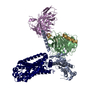 9d0aC 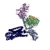 9e7rC M: atomic model generated by this map C: citing same article ( |
|---|---|
| Similar structure data |
-Links
| EMDB pages | EMDB (EBI/PDBe) / EMDataResource |
|---|---|
| Related items in Molecule of the Month |


-Map
| File | Download / File: emd_46571.map.gz / Format: CCP4 / Size: 52.7 MB / Type: IMAGE STORED AS FLOATING POINT NUMBER (4 BYTES) | ||||||||||||||||||||||||||||||||||||
|---|---|---|---|---|---|---|---|---|---|---|---|---|---|---|---|---|---|---|---|---|---|---|---|---|---|---|---|---|---|---|---|---|---|---|---|---|---|
| Projections & slices | Image control
Images are generated by Spider. | ||||||||||||||||||||||||||||||||||||
| Voxel size | X=Y=Z: 1.035 Å | ||||||||||||||||||||||||||||||||||||
| Density |
| ||||||||||||||||||||||||||||||||||||
| Symmetry | Space group: 1 | ||||||||||||||||||||||||||||||||||||
| Details | EMDB XML:
|
 Z (Sec.)
 Y (Row.)
 X (Col.)


-Supplemental data
-Half map: #2
| File | emd_46571_half_map_1.map | ||||||||||||
|---|---|---|---|---|---|---|---|---|---|---|---|---|---|
| Projections & Slices |
| ||||||||||||
| Density Histograms |


-Half map: #1
| File | emd_46571_half_map_2.map | ||||||||||||
|---|---|---|---|---|---|---|---|---|---|---|---|---|---|
| Projections & Slices |
| ||||||||||||
| Density Histograms |


- Sample components
-Entire : Complex of PAR1 with G proteins
| Entire | Name: Complex of PAR1 with G proteins |
|---|---|
| Components |
|
-Supramolecule #1: Complex of PAR1 with G proteins
| Supramolecule | Name: Complex of PAR1 with G proteins / type: complex / ID: 1 / Parent: 0 / Macromolecule list: #3, #1-#2, #4-#5 |
|---|---|
| Source (natural) | Organism: Homo sapiens (human) |
-Macromolecule #1: Guanine nucleotide-binding protein G(I)/G(S)/G(T) subunit beta-1
| Macromolecule | Name: Guanine nucleotide-binding protein G(I)/G(S)/G(T) subunit beta-1 type: protein_or_peptide / ID: 1 / Number of copies: 1 / Enantiomer: LEVO |
|---|---|
| Source (natural) | Organism: Homo sapiens (human) |
| Molecular weight | Theoretical: 37.285734 KDa |
| Recombinant expression | Organism:   Spodoptera frugiperda (fall armyworm) Spodoptera frugiperda (fall armyworm) |
| Sequence | String: SELDQLRQEA EQLKNQIRDA RKACADATLS QITNNIDPVG RIQMRTRRTL RGHLAKIYAM HWGTDSRLLV SASQDGKLII WDSYTTNKV HAIPLRSSWV MTCAYAPSGN YVACGGLDNI CSIYNLKTRE GNVRVSRELA GHTGYLSCCR FLDDNQIVTS S GDTTCALW ...String: SELDQLRQEA EQLKNQIRDA RKACADATLS QITNNIDPVG RIQMRTRRTL RGHLAKIYAM HWGTDSRLLV SASQDGKLII WDSYTTNKV HAIPLRSSWV MTCAYAPSGN YVACGGLDNI CSIYNLKTRE GNVRVSRELA GHTGYLSCCR FLDDNQIVTS S GDTTCALW DIETGQQTTT FTGHTGDVMS LSLAPDTRLF VSGACDASAK LWDVREGMCR QTFTGHESDI NAICFFPNGN AF ATGSDDA TCRLFDLRAD QELMTYSHDN IICGITSVSF SKSGRLLLAG YDDFNCNVWD ALKADRAGVL AGHDNRVSCL GVT DDGMAV ATGSWDSFLK IWN UniProtKB: Guanine nucleotide-binding protein G(I)/G(S)/G(T) subunit beta-1 |
-Macromolecule #2: Guanine nucleotide-binding protein G(I)/G(S)/G(O) subunit gamma-2
| Macromolecule | Name: Guanine nucleotide-binding protein G(I)/G(S)/G(O) subunit gamma-2 type: protein_or_peptide / ID: 2 / Number of copies: 1 / Enantiomer: LEVO |
|---|---|
| Source (natural) | Organism: Homo sapiens (human) |
| Molecular weight | Theoretical: 6.504446 KDa |
| Recombinant expression | Organism: Spodoptera frugiperda (fall armyworm) |
| Sequence | String: NTASIAQARK LVEQLKMEAN IDRIKVSKAA ADLMAYCEAH AKEDPLLTPV PASENPFRE UniProtKB: Guanine nucleotide-binding protein G(I)/G(S)/G(O) subunit gamma-2 |
-Macromolecule #3: Proteinase-activated receptor 1
| Macromolecule | Name: Proteinase-activated receptor 1 / type: protein_or_peptide / ID: 3 / Number of copies: 1 / Enantiomer: LEVO |
|---|---|
| Source (natural) | Organism: Homo sapiens (human) |
| Molecular weight | Theoretical: 43.020996 KDa |
| Recombinant expression | Organism: Spodoptera frugiperda (fall armyworm) |
| Sequence | String: SFLLRNPNDK YEPFWEDEEK NESGLTEYRL VSINKSSPLQ KQLPAFISED ASGYLTSSWL TLFVPSVYTG VFVVSLPLNI MAIVVFILK MKVKKPAVVY MLHLATADVL FVSVLPFKIS YYFSGSDWQF GSELCRFVTA AFYCNMYASI LLMTVISIDR F LAVVYPMQ ...String: SFLLRNPNDK YEPFWEDEEK NESGLTEYRL VSINKSSPLQ KQLPAFISED ASGYLTSSWL TLFVPSVYTG VFVVSLPLNI MAIVVFILK MKVKKPAVVY MLHLATADVL FVSVLPFKIS YYFSGSDWQF GSELCRFVTA AFYCNMYASI LLMTVISIDR F LAVVYPMQ SLSWRTLGRA SFTCLAIWAL AIAGVVPLLL KEQTIQVPGL NITTCHDVLN ETLLEGYYAY YFSAFSAVFF FV PLIISTV CYVSIIRCLS SSAVANRSKK SRALFLSAAV FCIFIICFGP TNVLLIAHYS FLSHTSTTEA AYFAYLLCVC VSS ISCCID PLIYYYASSE CQRYVYSILC CKESSDPSSY NSSGQLMASK MDTCSSNLNN SIYKKLLT UniProtKB: Proteinase-activated receptor 1 |
-Macromolecule #4: scFv16
| Macromolecule | Name: scFv16 / type: protein_or_peptide / ID: 4 / Number of copies: 1 / Enantiomer: LEVO |
|---|---|
| Source (natural) | Organism: |
| Molecular weight | Theoretical: 26.679721 KDa |
| Recombinant expression | Organism: Spodoptera frugiperda (fall armyworm) |
| Sequence | String: DVQLVESGGG LVQPGGSRKL SCSASGFAFS SFGMHWVRQA PEKGLEWVAY ISSGSGTIYY ADTVKGRFTI SRDDPKNTLF LQMTSLRSE DTAMYYCVRS IYYYGSSPFD FWGQGTTLTV SSGGGGSGGG GSGGGGSDIV MTQATSSVPV TPGESVSISC R SSKSLLHS ...String: DVQLVESGGG LVQPGGSRKL SCSASGFAFS SFGMHWVRQA PEKGLEWVAY ISSGSGTIYY ADTVKGRFTI SRDDPKNTLF LQMTSLRSE DTAMYYCVRS IYYYGSSPFD FWGQGTTLTV SSGGGGSGGG GSGGGGSDIV MTQATSSVPV TPGESVSISC R SSKSLLHS NGNTYLYWFL QRPGQSPQLL IYRMSNLASG VPDRFSGSGS GTAFTLTISR LEAEDVGVYY CMQHLEYPLT FG AGTKLEL KAAA |
-Macromolecule #5: Guanine nucleotide-binding protein G(q) subunit alpha chimera
| Macromolecule | Name: Guanine nucleotide-binding protein G(q) subunit alpha chimera type: protein_or_peptide / ID: 5 / Number of copies: 1 / Enantiomer: LEVO |
|---|---|
| Source (natural) | Organism: Homo sapiens (human) |
| Molecular weight | Theoretical: 41.972578 KDa |
| Recombinant expression | Organism: Spodoptera frugiperda (fall armyworm) |
| Sequence | String: MGCTLSAEDK AAVERSKMID RNLREDGEKA RRELKLLLLG TGESGKSTFI KQMRIIHGSG YSDEDKRGFT KLVYQNIFTA MQAMIRAMD TLKIPYKYEH NKAHAQLVRE VDVEKVSAFE NPYVDAIKSL WNDPGIQECY DRRREYQLSD STKYYLNDLD R VADPAYLP ...String: MGCTLSAEDK AAVERSKMID RNLREDGEKA RRELKLLLLG TGESGKSTFI KQMRIIHGSG YSDEDKRGFT KLVYQNIFTA MQAMIRAMD TLKIPYKYEH NKAHAQLVRE VDVEKVSAFE NPYVDAIKSL WNDPGIQECY DRRREYQLSD STKYYLNDLD R VADPAYLP TQQDVLRVRV PTTGIIEYPF DLQKVNFHMF DVGGQRSERR KWIQCFNDVT AIIFVVDSSD YNRLQEALND FK SIWNNRW LRTISVILFL NKQDLLAEKV LAGKSKIEDY FPEFARYTTP EDATPEPGED PRVTRAKYFI RKEFVDISTA SGD GRHICY PHFTCAVDTE NARRIFNDCK DIILQMNLRE YNLV |
-Experimental details
-Structure determination
| Method | cryo EM |
|---|---|
 Processing Processing | single particle reconstruction |
| Aggregation state | particle |
-Sample preparation
| Buffer | pH: 7 |
|---|---|
| Grid | Model: Quantifoil / Material: GOLD / Mesh: 300 / Support film - Material: GOLD / Support film - topology: HOLEY / Support film - Film thickness: 50 / Pretreatment - Type: GLOW DISCHARGE / Pretreatment - Time: 60 sec. |
| Vitrification | Cryogen name: ETHANE |
- Electron microscopy
| Microscope | TFS GLACIOS |
|---|---|
| Image recording | Film or detector model: TFS FALCON 4i (4k x 4k) / Average electron dose: 45.0 e/Å2 |
| Electron beam | Acceleration voltage: 200 kV / Electron source:  FIELD EMISSION GUN FIELD EMISSION GUN |
| Electron optics | Illumination mode: FLOOD BEAM / Imaging mode: BRIGHT FIELD / Nominal defocus max: 2.0 µm / Nominal defocus min: 0.8 µm |

