 Movie
Movie Controller

[English] 日本語
 Yorodumi
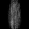
- PDB-9czl: Straight tau filaments in dominantly inherited Alzheimer disease ... -
+ Open data

- Basic information
| Entry | Database: PDB / ID: 9czl | |||||||||
|---|---|---|---|---|---|---|---|---|---|---|
| Title | Straight tau filaments in dominantly inherited Alzheimer disease with cotton wool plaques | |||||||||
 Components Components | Isoform Tau-C of Microtubule-associated protein tau | |||||||||
 Keywords Keywords | NEUROPEPTIDE / Tau filaments / SF / cotton wool plaques / neurodegeneration / PROTEIN FIBRIL | |||||||||
| Function / homology |  Function and homology information Function and homology informationplus-end-directed organelle transport along microtubule / histone-dependent DNA binding / negative regulation of protein localization to mitochondrion / neurofibrillary tangle / microtubule lateral binding / axonal transport / tubulin complex / positive regulation of protein localization to synapse / phosphatidylinositol bisphosphate binding / generation of neurons ...plus-end-directed organelle transport along microtubule / histone-dependent DNA binding / negative regulation of protein localization to mitochondrion / neurofibrillary tangle / microtubule lateral binding / axonal transport / tubulin complex / positive regulation of protein localization to synapse / phosphatidylinositol bisphosphate binding / generation of neurons / rRNA metabolic process / axonal transport of mitochondrion / regulation of mitochondrial fission / axon development / regulation of microtubule-based movement / intracellular distribution of mitochondria / regulation of chromosome organization / central nervous system neuron development / minor groove of adenine-thymine-rich DNA binding / lipoprotein particle binding / microtubule polymerization / negative regulation of mitochondrial membrane potential / regulation of microtubule polymerization / dynactin binding / apolipoprotein binding / protein polymerization / main axon / Caspase-mediated cleavage of cytoskeletal proteins / regulation of microtubule polymerization or depolymerization / negative regulation of mitochondrial fission / axolemma / glial cell projection / neurofibrillary tangle assembly / positive regulation of axon extension / regulation of cellular response to heat / positive regulation of microtubule polymerization / positive regulation of protein localization / Activation of AMPK downstream of NMDARs / positive regulation of superoxide anion generation / cellular response to brain-derived neurotrophic factor stimulus / regulation of long-term synaptic depression / supramolecular fiber organization / cytoplasmic microtubule organization / regulation of calcium-mediated signaling / axon cytoplasm / somatodendritic compartment / synapse assembly / phosphatidylinositol binding / nuclear periphery / astrocyte activation / enzyme inhibitor activity / protein phosphatase 2A binding / stress granule assembly / regulation of microtubule cytoskeleton organization / regulation of autophagy / cellular response to reactive oxygen species / microglial cell activation / cellular response to nerve growth factor stimulus / Hsp90 protein binding / protein homooligomerization / SH3 domain binding / PKR-mediated signaling / synapse organization / regulation of synaptic plasticity / response to lead ion / microtubule cytoskeleton organization / memory / neuron projection development / cytoplasmic ribonucleoprotein granule / cell-cell signaling / single-stranded DNA binding / cellular response to heat / protein-folding chaperone binding / growth cone / microtubule cytoskeleton / actin binding / cell body / double-stranded DNA binding / microtubule binding / sequence-specific DNA binding / amyloid fibril formation / dendritic spine / microtubule / protein-macromolecule adaptor activity / learning or memory / neuron projection / membrane raft / negative regulation of gene expression / axon / neuronal cell body / DNA damage response / dendrite / protein kinase binding / enzyme binding / mitochondrion / DNA binding / RNA binding / extracellular region / identical protein binding / nucleus Similarity search - Function | |||||||||
| Biological species |  Homo sapiens (human) Homo sapiens (human) | |||||||||
| Method | ELECTRON MICROSCOPY / helical reconstruction / cryo EM / Resolution: 2.9 Å | |||||||||
 Authors Authors | Hoq, M.R. / Vago, F.S. / Ozcan, K.A. / Bharath, S.R. | |||||||||
| Funding support |  United States, 2items United States, 2items
| |||||||||
 Citation Citation | Journal: Acta Neuropathol / Year: 2024 Title: Cryo-EM structures of cotton wool plaques' amyloid β and of tau filaments in dominantly inherited Alzheimer disease. Authors: Md Rejaul Hoq / Anllely Fernandez / Frank S Vago / Grace I Hallinan / Sakshibeedu R Bharath / Daoyi Li / Kadir A Ozcan / Holly J Garringer / Wen Jiang / Ruben Vidal / Bernardino Ghetti / Abstract: Cotton wool plaques (CWPs) have been described as features of the neuropathologic phenotype of dominantly inherited Alzheimer disease (DIAD) caused by some missense and deletion mutations in the ...Cotton wool plaques (CWPs) have been described as features of the neuropathologic phenotype of dominantly inherited Alzheimer disease (DIAD) caused by some missense and deletion mutations in the presenilin 1 (PSEN1) gene. CWPs are round, eosinophilic amyloid-β (Aβ) plaques that lack an amyloid core and are recognizable, but not fluorescent, in Thioflavin S (ThS) preparations. Amino-terminally truncated and post-translationally modified Aβ peptide species are the main component of CWPs. Tau immunopositive neurites may be present in CWPs. In addition, neurofibrillary tangles coexist with CWPs. Herein, we report the structure of Aβ and tau filaments isolated from brain tissue of individuals affected by DIAD caused by the PSEN1 V261I and A431E mutations, with the CWP neuropathologic phenotype. CWPs are predominantly composed of type I Aβ filaments present in two novel arrangements, type Ic and type Id; additionally, CWPs contain type I and type Ib Aβ filaments. Tau filaments have the AD fold, which has been previously reported in sporadic AD and DIAD. The formation of type Ic and type Id Aβ filaments may be the basis for the phenotype of CWPs. Our data are relevant for the development of PET imaging methodologies to best detect CWPs in DIAD. | |||||||||
| History |
|
- Structure visualization
| Structure viewer | Molecule: MolmilJmol/JSmol |
|---|
- Downloads & links
-Download
| PDBx/mmCIF format | 9czl.cif.gz | 136.7 KB | Display | PDBx/mmCIF format |
|---|---|---|---|---|
| PDB format | pdb9czl.ent.gz | 110 KB | Display | PDB format |
| PDBx/mmJSON format | 9czl.json.gz | Tree view | PDBx/mmJSON format | |
| Others |  Other downloads Other downloads |
-Validation report
| Arichive directory | https://data.pdbj.org/pub/pdb/validation_reports/cz/9czlftp://data.pdbj.org/pub/pdb/validation_reports/cz/9czl | HTTPS FTP |
|---|
-Related structure data
| Related structure data | 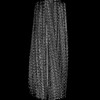 46420MC  9cziC 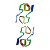 9cznC 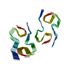 9czpC M: map data used to model this data C: citing same article ( |
|---|---|
| Similar structure data |
-Links
 PDBj
PDBj


- Assembly
| Deposited unit | 
|
|---|---|
| 1 |
|
-Components
| #1: Protein | Mass: 8069.321 Da / Num. of mol.: 10 / Fragment: UNP residues 274-347 / Source method: isolated from a natural source / Source: (natural) Homo sapiens (human) / References: UniProt: P10636 |
|---|
-Experimental details
-Experiment
| Experiment | Method: ELECTRON MICROSCOPY |
|---|---|
| EM experiment | Aggregation state: FILAMENT / 3D reconstruction method: helical reconstruction |
- Sample preparation
| Component | Name: Straight tau filament / Type: TISSUE / Entity ID: all / Source: NATURAL | |||||||||||||||
|---|---|---|---|---|---|---|---|---|---|---|---|---|---|---|---|---|
| Source (natural) | Organism: Homo sapiens (human) | |||||||||||||||
| Buffer solution | pH: 7.4 | |||||||||||||||
| Buffer component |
| |||||||||||||||
| Specimen | Embedding applied: NO / Shadowing applied: NO / Staining applied: NO / Vitrification applied: YES | |||||||||||||||
| Vitrification | Instrument: GATAN CRYOPLUNGE 3 / Cryogen name: ETHANE |
- Electron microscopy imaging
| Experimental equipment |  Model: Titan Krios / Image courtesy: FEI Company |
|---|---|
| Microscopy | Model: FEI TITAN KRIOS |
| Electron gun | Electron source:  FIELD EMISSION GUN / Accelerating voltage: 300 kV / Illumination mode: FLOOD BEAM FIELD EMISSION GUN / Accelerating voltage: 300 kV / Illumination mode: FLOOD BEAM |
| Electron lens | Mode: BRIGHT FIELD / Nominal magnification: 81000 X / Nominal defocus max: 2500 nm / Nominal defocus min: 500 nm / Cs: 2.7 mm / C2 aperture diameter: 50 µm |
| Specimen holder | Cryogen: NITROGEN / Specimen holder model: FEI TITAN KRIOS AUTOGRID HOLDER |
| Image recording | Average exposure time: 3.21 sec. / Electron dose: 57.79 e/Å2 / Detector mode: COUNTING / Film or detector model: GATAN K3 BIOQUANTUM (6k x 4k) / Num. of grids imaged: 1 / Num. of real images: 17581 |
| EM imaging optics | Energyfilter name: GIF Bioquantum / Energyfilter slit width: 20 eV |
| Image scans | Sampling size: 5 µm / Width: 5760 / Height: 4092 |
- Processing
| EM software |
| ||||||||||||||||||||||||||||||||||||||||||||
|---|---|---|---|---|---|---|---|---|---|---|---|---|---|---|---|---|---|---|---|---|---|---|---|---|---|---|---|---|---|---|---|---|---|---|---|---|---|---|---|---|---|---|---|---|---|
| CTF correction | Type: PHASE FLIPPING AND AMPLITUDE CORRECTION | ||||||||||||||||||||||||||||||||||||||||||||
| Helical symmerty | Angular rotation/subunit: -1.08 ° / Axial rise/subunit: 4.78 Å / Axial symmetry: C1 | ||||||||||||||||||||||||||||||||||||||||||||
| 3D reconstruction | Resolution: 2.9 Å / Resolution method: FSC 0.143 CUT-OFF / Num. of particles: 24819 / Symmetry type: HELICAL | ||||||||||||||||||||||||||||||||||||||||||||
| Atomic model building | Protocol: AB INITIO MODEL / Space: REAL |
