 Movie
Movie Controller


+ Open data

- Basic information
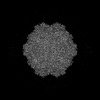
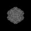
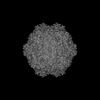
| Entry | Database: PDB / ID: 9cuz | ||||||
|---|---|---|---|---|---|---|---|
| Title | Bufavirus 1 complexed with 6SLN | ||||||
 Components Components | VP1 | ||||||
 Keywords Keywords | VIRUS LIKE PARTICLE / Bufavirus / parvovirus / glycan | ||||||
| Function / homology |  Function and homology information Function and homology information | ||||||
| Biological species |  Bufavirus-1 Bufavirus-1 | ||||||
| Method | ELECTRON MICROSCOPY / single particle reconstruction / cryo EM / Resolution: 2.16 Å | ||||||
 Authors Authors | Gulkis, M.C. / McKenna, R. / Bennett, A.D. | ||||||
| Funding support |  United States, 1items United States, 1items
| ||||||
 Citation Citation | Journal: Viruses / Year: 2024 Title: Structural Characterization of Human Bufavirus 1: Receptor Binding and Endosomal pH-Induced Changes. Authors: Mitchell Gulkis / Mengxiao Luo / Paul Chipman / Mario Mietzsch / Maria Söderlund-Venermo / Antonette Bennett / Robert McKenna /  Abstract: Bufaviruses (BuV) are members of the of the genus. They are non-enveloped, T = 1 icosahedral ssDNA viruses isolated from patients exhibiting acute diarrhea. The lack of treatment options and a ...Bufaviruses (BuV) are members of the of the genus. They are non-enveloped, T = 1 icosahedral ssDNA viruses isolated from patients exhibiting acute diarrhea. The lack of treatment options and a limited understanding of their disease mechanisms require studying these viruses on a molecular and structural level. In the present study, we utilize glycan arrays and cell binding assays to demonstrate that BuV1 capsid binds terminal sialic acid (SIA) glycans. Furthermore, using cryo-electron microscopy (cryo-EM), SIA is shown to bind on the 2/5-fold wall of the capsid surface. Interestingly, the capsid residues stabilizing SIA binding are conserved in all human BuVs identified to date. Additionally, biophysical assays illustrate BuV1 capsid stabilization during endo-lysosomal (pH 7.4-pH 4) trafficking and capsid destabilization at pH 3 and less, which correspond to the pH of the stomach. Hence, we determined the cryo-EM structures of BuV1 capsids at pH 7.4, 4.0, and 2.6 to 2.8 Å, 3.2 Å, and 2.7 Å, respectively. These structures reveal capsid structural rearrangements during endo-lysosomal escape and provide a potential mechanism for this process. The structural insights gained from this study will add to the general knowledge of human pathogenic parvoviruses. Furthermore, the identification of the conserved SIA receptor binding site among BuVs provides a possible targetable surface-accessible pocket for the design of small molecules to be developed as anti-virals for these viruses. | ||||||
| History |
|
- Structure visualization
| Structure viewer | Molecule: MolmilJmol/JSmol |
|---|
- Downloads & links
-Download
| PDBx/mmCIF format | 9cuz.cif.gz | 6.1 MB | Display | PDBx/mmCIF format |
|---|---|---|---|---|
| PDB format | pdb9cuz.ent.gz | Display | PDB format | |
| PDBx/mmJSON format | 9cuz.json.gz | Tree view | PDBx/mmJSON format | |
| Others |  Other downloads Other downloads |
-Validation report
| Arichive directory | https://data.pdbj.org/pub/pdb/validation_reports/cu/9cuzftp://data.pdbj.org/pub/pdb/validation_reports/cu/9cuz | HTTPS FTP |
|---|
-Related structure data
| Related structure data | 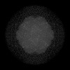 45954MC 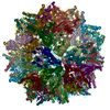 9cv0C 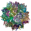 9cv9C 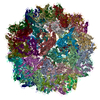 9cwsC M: map data used to model this data C: citing same article ( |
|---|---|
| Similar structure data |
-Links
 PDBj
PDBj


- Assembly
| Deposited unit | 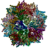
|
|---|---|
| 1 |
|
-Components
| #1: Protein | Mass: 80543.203 Da / Num. of mol.: 60 Source method: isolated from a genetically manipulated source Source: (gene. exp.) Bufavirus-1 / Gene: BF-002 / Cell line (production host): Sf9 / Production host:   Spodoptera frugiperda (fall armyworm) / References: UniProt: A0A097PIM0 Spodoptera frugiperda (fall armyworm) / References: UniProt: A0A097PIM0#2: Sugar | ChemComp-SIA /   Type: D-saccharide, alpha linking / Mass: 309.270 Da / Num. of mol.: 60 Type: D-saccharide, alpha linking / Mass: 309.270 Da / Num. of mol.: 60Source method: isolated from a genetically manipulated source Formula: C11H19NO9 / Feature type: SUBJECT OF INVESTIGATION Has ligand of interest | Y | |
|---|
-Experimental details
-Experiment
| Experiment | Method: ELECTRON MICROSCOPY |
|---|---|
| EM experiment | Aggregation state: PARTICLE / 3D reconstruction method: single particle reconstruction |
- Sample preparation
| Component | Name: Bufavirus-1 / Type: VIRUS / Details: Bufavirus 1 VP2 was overexpressed in Sf9 cells / Entity ID: #1 / Source: RECOMBINANT | |||||||||||||||||||||||||
|---|---|---|---|---|---|---|---|---|---|---|---|---|---|---|---|---|---|---|---|---|---|---|---|---|---|---|
| Molecular weight | Value: 4 MDa / Experimental value: NO | |||||||||||||||||||||||||
| Source (natural) | Organism: Bufavirus-1 | |||||||||||||||||||||||||
| Source (recombinant) | Organism: Spodoptera frugiperda (fall armyworm) | |||||||||||||||||||||||||
| Details of virus | Empty: YES / Enveloped: NO / Isolate: STRAIN / Type: VIRUS-LIKE PARTICLE | |||||||||||||||||||||||||
| Natural host | Organism: Homo sapiens | |||||||||||||||||||||||||
| Virus shell | Diameter: 260 nm / Triangulation number (T number): 1 | |||||||||||||||||||||||||
| Buffer solution | pH: 7.4 | |||||||||||||||||||||||||
| Buffer component |
| |||||||||||||||||||||||||
| Specimen | Conc.: 0.5 mg/ml / Embedding applied: NO / Shadowing applied: NO / Staining applied: NO / Vitrification applied: YES | |||||||||||||||||||||||||
| Specimen support | Grid material: COPPER / Grid type: Quantifoil R2/2 | |||||||||||||||||||||||||
| Vitrification | Instrument: FEI VITROBOT MARK IV / Cryogen name: ETHANE |
- Electron microscopy imaging
| Experimental equipment |  Model: Titan Krios / Image courtesy: FEI Company |
|---|---|
| Microscopy | Model: TFS KRIOS |
| Electron gun | Electron source:  FIELD EMISSION GUN / Accelerating voltage: 300 kV / Illumination mode: FLOOD BEAM FIELD EMISSION GUN / Accelerating voltage: 300 kV / Illumination mode: FLOOD BEAM |
| Electron lens | Mode: BRIGHT FIELD / Nominal defocus max: 3600 nm / Nominal defocus min: 100 nm / Cs: 2.7 mm |
| Specimen holder | Cryogen: NITROGEN |
| Image recording | Electron dose: 75 e/Å2 / Detector mode: COUNTING / Film or detector model: GATAN K2 SUMMIT (4k x 4k) / Num. of real images: 1115 |
| EM imaging optics | Energyfilter slit width: 20 eV |
| Image scans | Movie frames/image: 50 / Used frames/image: 1-50 |
- Processing
| EM software | Name: PHENIX / Version: 1.21_5207: / Category: model refinement |
|---|---|
| CTF correction | Type: PHASE FLIPPING AND AMPLITUDE CORRECTION |
| Particle selection | Num. of particles selected: 250990 |
| Symmetry | Point symmetry: I (icosahedral) |
| 3D reconstruction | Resolution: 2.16 Å / Resolution method: FSC 0.143 CUT-OFF / Num. of particles: 95075 / Num. of class averages: 1 / Symmetry type: POINT |
| Atomic model building | B value: 90.4 / Protocol: RIGID BODY FIT / Space: REAL / Target criteria: Cross-correlation coefficient |
| Atomic model building | PDB-ID: 6BWX Accession code: 6BWX / Source name: PDB / Type: experimental model |

